Aldehyde Stabilized Cryopreserved Pig Brain Evaluation Images
This page contains a subset of the electron microscopic imaging that was performed on the 21st Century Medicine / Robert McIntyre pig brain entry into the Brain Preservation Foundation (BPF) Brain Preservation Technology Prize.
The Pig brain was preserved using the Aldehyde Stabilized Cryopreservation (ASC) technique as described in the 2015 Cryobiology paper “Aldehyde Stabilized Cryopreservation” by Robert McIntyre and Gregory Fahy. The paper is open access and can be downloaded at the following link:
https://doi.org/10.1016/j.cryobiol.2015.09.003
The requirements of the Brain Preservation Technology Prize state that the connectome (the pattern of synaptic connectivity) of a whole large mammal brain must be preserved in a fashion that would allow for extremely long-term storage (>100 years). As part of this evaluation, a representative from the BPF (Kenneth Hayworth) visited the 21st Century Medicine (21CM) laboratory in Fontana California. He was present for the rewarming of this pig brain as it was removed from -135o C cold storage. The BPF representative witnessed that the brain was ‘frozen’ solid (more properly it was vitrified solid) and witnessed it rewarming to room temperature. The BPF representative witnessed the dissection of the brain first into hemispheres and then into chunks of suitable size for vibratomy. Pictures of this rewarming and dissection are shown in Figure 1.
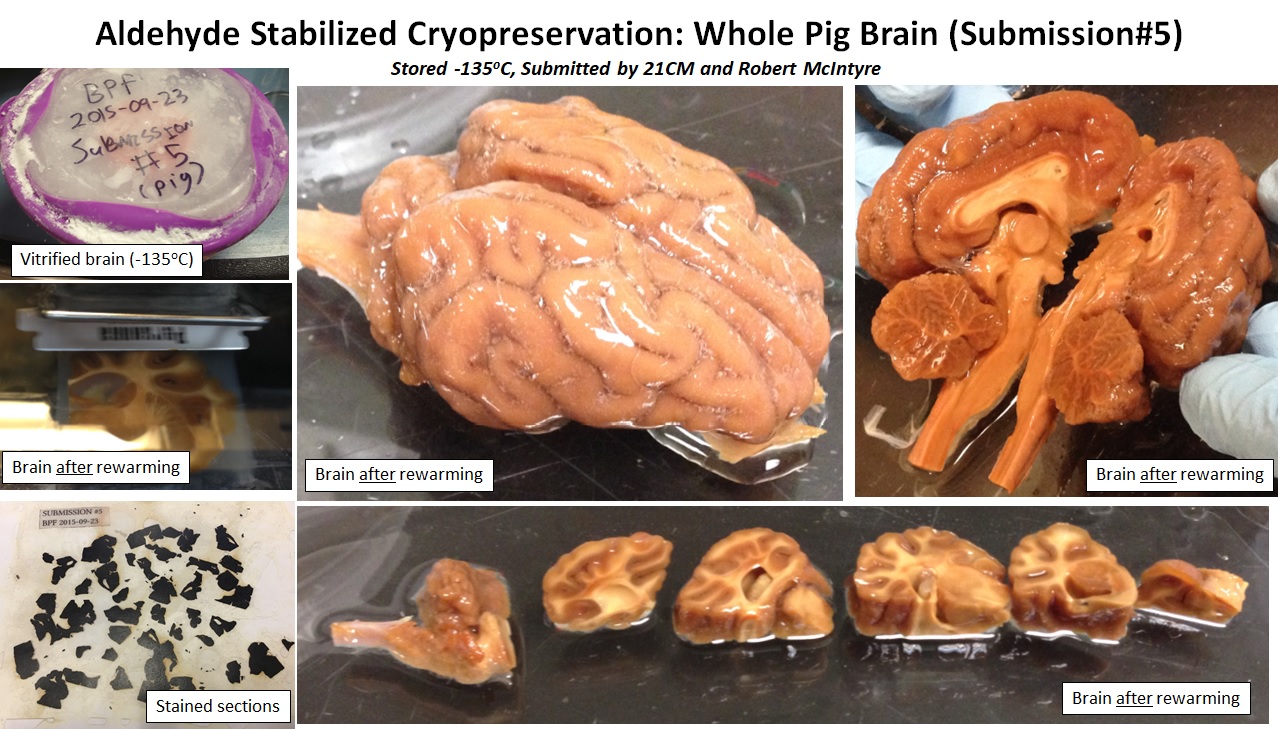 Figure 1. Images of ASC-preserved pig brain dissection.
The BPF representative observed this dissection closely looking for any macroscopic signs of ice crystal damage. None were observed. Then several of the tissue blocks were vibratomed at 100 to 200 micron thickness. This again offered the chance to look for macroscopic damage. Again none was observed. Finally several vibratome sections were taken from each of four tissue blocks and put through a standard chemical process to prepare them for 3D electron microscopy (i.e. osmium fixation, en bloc heavy metal staining, dehydration, resin infiltration, and plastic embedding). The BPF representative then left the 21CM facility taking with him these plastic-embedded pig brain sections for further off-site electron microscopic evaluation.
One pseudo-randomly chosen location in each of four different vibratome sections was chosen for Focused Ion Beam Scanning Electron Microscopy (FIB-SEM). FIB-SEM images of several other regions were also performed and all gave results similar to these presented. In general the ultrastructure of synaptic junctions seemed well preserved across all of the tested regions—equivalent to other glutaraldehyde perfused mammalian samples used in connectomics research studies. Neural processes appear traceable. The most significant artifacts observed were ‘myelin figures’ (abnormal congregations of lipids within cells which are thought to be an artifact of lipid fixation). These artifacts do not appear to significantly impact connectome traceability in the FIB-SEM volumes.
Below are overview images of the FIB-SEM datasets. Clicking on each will bring you to a YouTube video that scans through the 3D FIB-SEM volume dataset one image plane at a time:
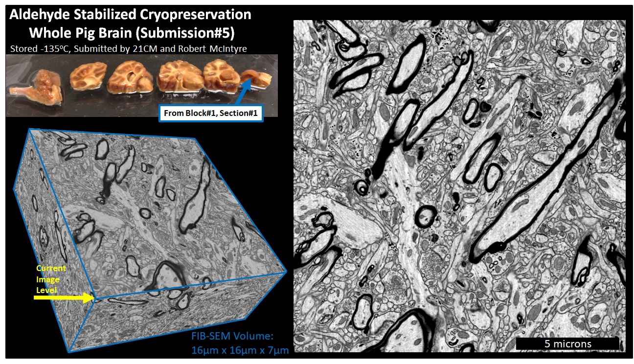
Figure 2. Overview of FIB-SEM volume of ASC-preserved pig brain acquired from Block#1 Section#1. Link to YouTube video , Link to 16nm Isotropic 3D Data File
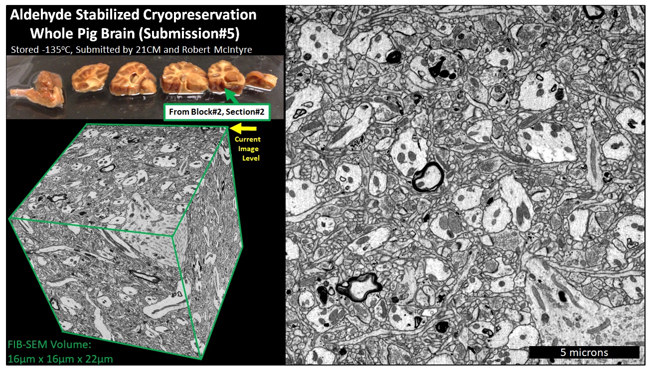
Figure 3. Overview of FIB-SEM volume of ASC-preserved pig brain acquired from Block#2 Section#2. Link to YouTube video , Link to 16nm Isotropic 3D Data File
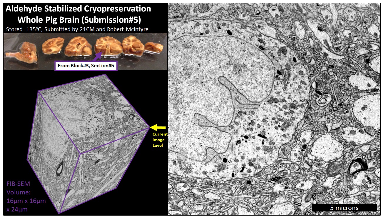 Figure 4. Overview of FIB-SEM volume of ASC-preserved pig brain acquired from Block#3 Section#5. Link to YouTube video , Link to 16nm Isotropic 3D Data File
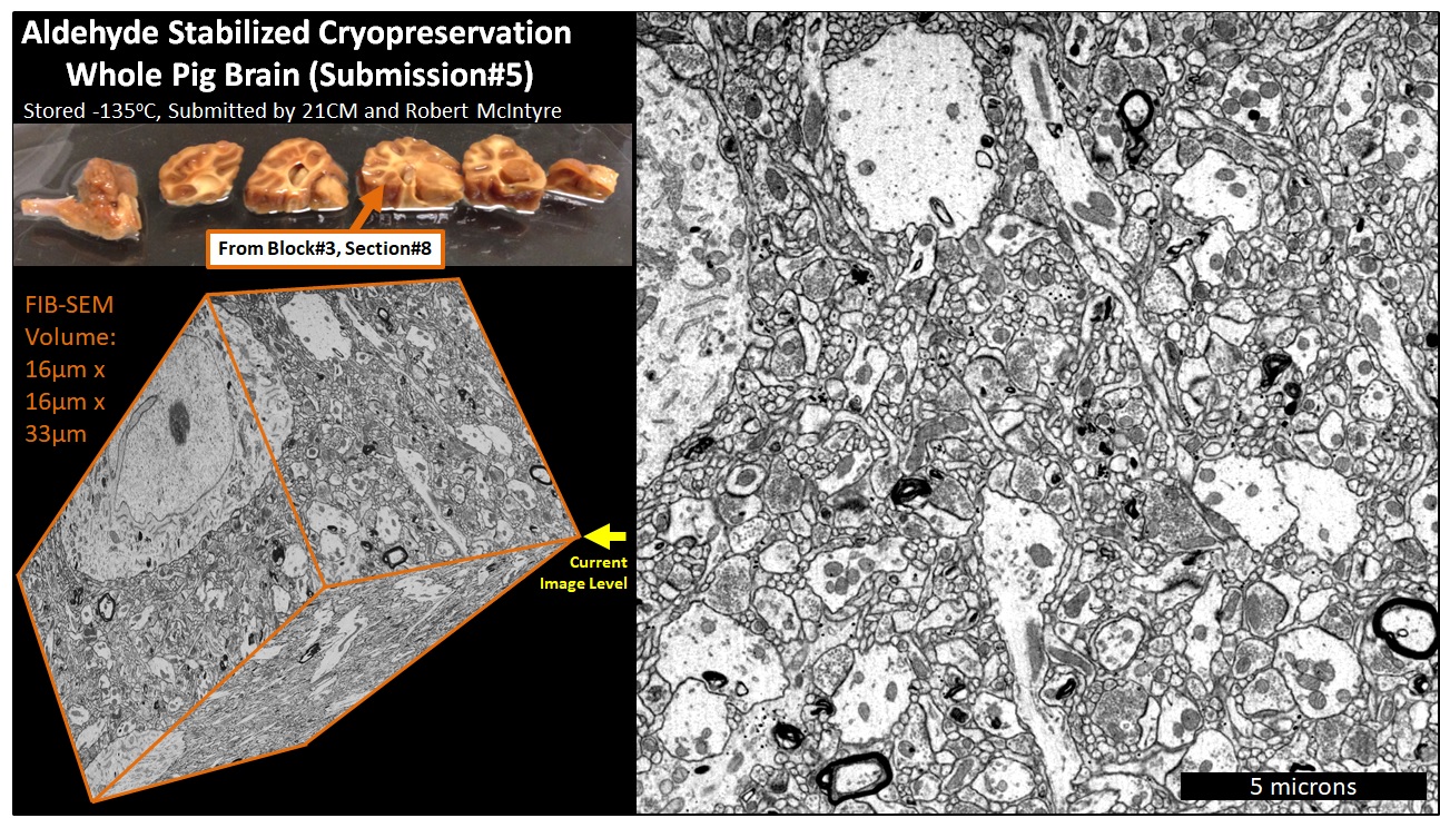 Figure 5. Overview of FIB-SEM volume of ASC-preserved pig brain acquired from Block#3 Section#8. Link to YouTube video , Link to 16nm Isotropic 3D Data File
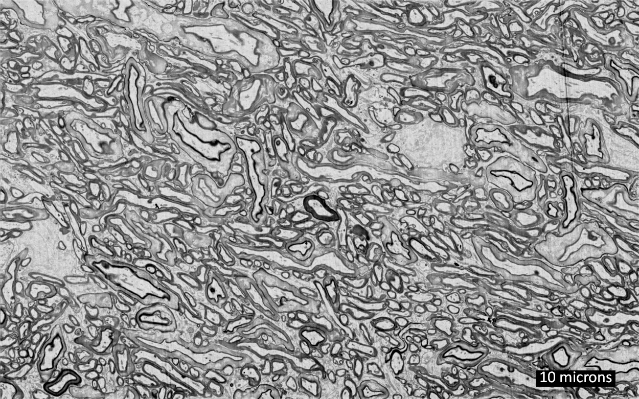
Figure 6. Representative electron micrograph of white matter region in ASC-preserved pig brain acquired from Block#3 Section#6.
The complete collection of FIB-SEM volumes can be obtained at the following Google Drive link:
CLICK HERE FOR GOOGLE DRIVE LINK TO DOWNLOAD 3D DATASETS
This link contains the following FIBSEM datasets:
- ASC_Pig_Submission5_Block1_Section1_16nmIso_16x16x7microns.avi
- ASC_Pig_Submission5_Block1_Section1_32nmIso_38x37x7microns.avi
- ASC_Pig_Submission5_Block2_Section2_16nmIso_16x16x22microns.avi
- ASC_Pig_Submission5_Block2_Section2_32nmIso_27x28x22microns.avi
- ASC_Pig_Submission5_Block3_Section5_16nmIso_16x16x24microns.avi
- ASC_Pig_Submission5_Block3_Section5_32nmIso_46x19x24microns.avi
- ASC_Pig_Submission5_Block3_Section8_16nmIso_16x16x33microns.avi
- ASC_Pig_Submission5_Block3_Section8_32nmIso_27x28x33microns.avi