
May 2015 BPF Prize Update
Executive Summary
Brain Preservation Prize competitor Shawn Mikula just published his whole mouse brain electron microscopy protocol in Nature Methods (paper, BPF interview), putting him close to winning the mouse phase of our prize.
Brain Preservation Prize competitor 21st Century Medicine has developed a new “Aldehyde-Stabilized Cryopreservation” technique–preliminary results show good ultrastructure preservation even after storage of a whole rabbit brain at -135 degrees C.
Update on Dr. Shawn Mikula Progress
A major milestone has just been reached in our Brain Preservation Prize. Shawn Mikula (Max Planck Institute for Medical Research in Heidelberg) has just published the first ever paper demonstrating how an entire mouse brain can be preserved at the ultrastructure level for electron microscopic (EM) imaging of its entire connectome. His paper “High-resolution whole-brain staining for electron microscopic circuit reconstruction” was published in the prestigious journal Nature Methods in an advanced online format last week. Here is a link.
Over the last few years I have interacted frequently with Dr. Mikula (both in my capacity as part of the Brain Preservation Foundation and as part of my own neuroscience research) and I have had the opportunity to image many of the mouse brains he has prepared as part of his protocol development work. However, I promised him not to publicly disclose the details of his protocol or the imaging results until after he had the opportunity to officially publish them in a peer-reviewed science journal. So I can now finally bring you all up to speed on what specifically he has accomplished and discuss how close he is to meeting the criteria for the mouse phase of our prize.
As is well known to all electron microscopists, the traditional protocol for preparing brain tissue for EM imaging only works for small pieces of tissue. The key problem has been that the chemicals used to preserve (and stain) the lipid membrane of cells, a mixture of osmium tetroxide with potassium ferrocyanide (OsO4 / K4Fe(CN)6), is prone to precipitation and barrier formation within the tissue. This has limited high-quality ultrastructure preservation and staining to depths of just a few hundred microns thick. Dr. Mikula’s paper shows that this can be overcome by adding a high concentration of formamide to the mix. According to his paper an aqueous solution containing 40 mM OsO4, 35 mM K4Fe(CN)6, 100 mM Cacodylate buffer (pH 7.4) and 2.5 M formamide is sufficient to completely eliminate barrier formation allowing for uniform preservation and staining of an entire mouse brain.
Dr. Mikula’s paper provides extensive EM and x-ray imaging data to back up this claim. Quoting from the abstract: “Here we describe a preparation, BROPA (brain-wide reduced-osmium staining with pyrogallol-mediated amplification), that results in the preservation and staining of ultrastructural details throughout the brain at a resolution necessary for tracing neuronal processes and identifying synaptic contacts between them. Using serial block-face electron microscopy (SBEM), we tested human annotator ability to follow neural ‘wires’ reliably and over long distances as well as the ability to detect synaptic contacts. Our results suggest that the BROPA method can produce a preparation suitable for the reconstruction of neural circuits spanning an entire mouse brain.”
Are these results sufficient for Mikula to win the mouse phase of our Brain Preservation Prize? The short answer is yes –if the claims made in the paper can be verified by our imaging then Mikula will be awarded the mouse phase of our prize. On two separate occasions in the past, Dr. Mikula has provide the BPF with a mouse brain for official evaluation, however on both of those occasions we found damage sufficient to reject the entry. The first brain we imaged showed cortical cracks when x-ray imaged. In the second brain, osmium penetration was not uniform leaving core regions like the thalamus insufficiently preserved. Dr. Mikula’s paper discusses the crack issue and its likely causes and solutions. He has said that he will be providing the BPF with a new official entry soon.
Another key question is whether his ‘formamide’ technique will be able to be scaled up to a large mammal –like the pig brain required for the final phase of our prize, or a human brain? Dr. Mikula is already working to procure high-quality glutaraldehyde perfused pig brains on which to test his technique. I suspect that to scale up to these large brains his protocol will need to be modified to include vascular perfusion of his osmium/formamide solution. And it may require the other steps of his process (dehydration and resin infiltration) to likewise be assisted by vascular perfusion.
Overall I think Dr. Mikula has overcome what is perhaps the key roadblock to whole brain EM preparation, and I think there is every reason to be optimistic that extension of his technique from mouse to human application is not only technically feasible but within reach. I very much hope that Dr. Mikula’s breakthrough publication will be recognized and followed up on not only within the neuroscience and connectomics fields, but also within the medical field as it brings us one small, but significant, step closer to an emergency medical procedure which could preserve a terminal patient’s brain for long-term storage with sufficient fidelity to preserve memory and identity.
Update on 21st Century Medicine Progress
I want to also touch on the significant progress that has been made by our other competitor team, 21st Century Medicine (21CM). Unfortunately issues of publication have also prevented me from disclosing many of the details of their research, but I can assure you that they have been hard at work competing in our prize–and they, like Mikula, have their sights set on eventually winning.
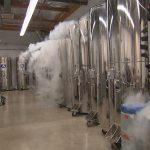
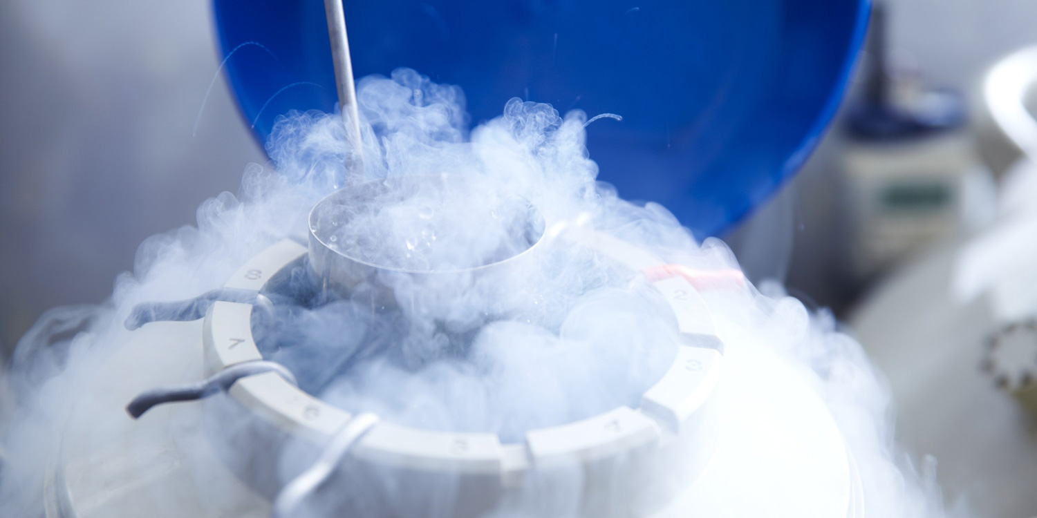
21CM’s core mission is to develop a cryopreservation protocol sufficiently benign that whole, donated human organs could be vitrified (stored below -130 degrees Celsius without ice formation) and rewarmed when needed for transplantation. They have had great success showing that viability can be restored in vitrified slices of tissue as shown in their paper, “Cryopreservation of precision-cut tissue slices.”
Figure 6 of that paper shows the preliminary results of experiments (more are due to be published) where living brain slices through the hippocampus of rats and rabbits were vitrified and stored for days to months at approximately -130 degrees Celsius. These tissue slices were then rewarmed, washed of cryoprotectant and tested for neuronal responses including Long Term Potentiation (LTP), the basis of memory formation in the hippocampus. Quoting from that paper:
“[The figure shows the] lack of effect of vitrification on the long term potentiation (LTP) response, a form of neurophysiological “memory” which consists of a permanent increase in the magnitude of the response to a given CA3 cell stimulation (recorded in this case as the amplitude of the excitatory post-synaptic field potentials at the Schaffer collateral-CA1 dendrite junction) as a result of prior “training” (intensive stimulation) of the involved synapses. Control brain slices increased their field EPSP response to about 30% above the baseline response amplitude (LTP ratio of about 1.3) in response to prior “training.” The same basic result was also seen after loading and unloading of VM3 (LU); after loading of VM3, vitrification, rewarming, and unloading of VM3 (VIT); and after storage of vitrified slices for days to months below the glass transition temperature (STR; storage time had no effect on the results obtained).”
With recovery-of-function results like this, one might expect that demonstrating whole brain ultrastructure preservation for our Brain Preservation Prize would be trivial. Unfortunately it is much easier to get cryoprotectant solutions into and out of half millimeter slices than whole brains. The whole rabbit brains that 21CM has perfused with cryoprotectant agents (CPA) for our prize have shown significant amounts of shrinkage due to dehydration from the high concentration and fast ramping of CPA used. Electron micrographs of this tissue are thus difficult to interpret and we have been unable to accurately assess the degree of ultrastructure preservation by this technique. 21CM has ideas on how to overcome this hurdle (which they believe to be one of evaluation rather than preservation) but progress has stalled on those experiments due to the expense involved.
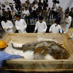
Recently however, 21CM has begun a set of experiments (spearheaded by recent MIT graduate Robert Louis McIntyre) which overcomes this dehydration and shrinkage issue in a very simple and inexpensive, but unorthodox, way. They perfuse the rabbit brain with glutaraldehyde fixative prior to perfusion with CPA and low temperature vitrification! This pre-fixation is of course completely incompatible with recovery of function by simple rewarming, but it has the effect of stabilizing the vascular system and tissue sufficiently to allow long duration room temperature perfusion of CPA. Initial results show that these brains (stored intact briefly at -135 degrees C) are not shrunken by this procedure and electron micrographs of brain ultrastructure appear “textbook-normal”. We have begun 3D EM imaging studies on samples provided by 21CM and our initial results show that connectomic details are indeed well preserved–the fine neural processes remain traceable in 3D and synaptic contacts are clearly seen. (It should be noted that the BPF had to put in the research funding necessary to support these experiments.)
How close is 21CM to winning our Brain Preservation Prize? Since a requirement of winning the prize (even the mouse phase) is publication of the protocol, it is clear that 21CM is behind Dr. Mikula in this regard. Also, Dr. Mikula has provided very extensive 3D EM evidence of connectome preservation across a whole brain, in contrast 21CM has so far only provided 2D electron micrographs showing good preservation of ultrastructure in the hippocampal region of vitrified whole rabbit brains. However, it could be argued that 21CM’s new technique (which could be called “Aldehyde-Stabilized Cryopreservation”) would be much more readily transitioned to a large mammal like a pig since all of its steps are already perfusion based. 21CM has even begun preliminary experiments applying their technique to whole pig brains. In this sense, Dr. Mikula is close to winning the mouse phase of our prize, but 21CM could very well be on their way to winning the final phase of our prize.
It should also be noted that this Aldehyde-Stabilized Cryopreservation technique has several potential advantages. Aldehyde-stabilized cryopreservation avoids the requirement of plastic embedding for long-term storage, meaning that, unlike Dr. Mikula’s protocol, no aggressive organic solvent step is necessary; lipids do not need to be further stabilized–thus there is no requirement for osmium, avoiding its expense and penetration issues entirely. But more importantly, this means that this aldehyde-stabilized cryopreservation would result in much fewer changes at the molecular level to brain tissue. Studies have long shown that although glutaraldehyde crosslinks proteins (stopping decay) it is quite good at preserving their sequential structure and positions within the cell.
Aldehyde-stabilized cryopreservation may even have advantages over an optimized cryopreservation protocol. The initial perfusion of glutaraldehyde is almost instantly effective in stopping cellular decay and locks crucial molecules like ion channels and receptor proteins in place. This allows the subsequent CPA perfusion to be optimized (in terms of concentrations, temperature, and timing) to achieve the best possible structural preservation of the brain’s connectome.
It should be noted that Aldehyde-Stabilized Cryopreservation has been suggested in the past. In fact its potential advantages were discussed extensively in chapter 9 of Eric Drexler’s 1987 book “Engines of Creation”.Here is an online link.
To our knowledge, 21CM’s recent experiments are the first and only serious attempt to test this Aldehyde-Stabilized Cryopreservation technique’s ability to preserve a whole mammalian brain.
In summary, I am very pleased to report that significant progress has been made by both of our competitor teams. Dr. Mikula’s recent publication shows that he is very close to meeting the requirements for the mouse phase of our prize, and both he and 21CM have presented plans for how they intend to meet the requirement of the large-mammal phase of our prize.
– Kenneth Hayworth
President of the Brain Preservation Foundation


