
The preservation of extracellular space in the brain
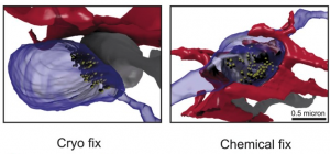
Cryo-fixed tissue has more space between the synapse and astrocytic processes than in chemically fixed tissue; http://www.ncbi.nlm.nih.gov/pmc/articles/PMC4530226/
Although most people usually focus on the brain cells when discussing brain preservation techniques, extracellular space is also worthy of consideration. In vivo, extracellular space makes up around 1/5th of overall brain volume, although this varies based on brain region, developmental stage, and surely many other factors. Over the past few months, Pallotto et al and Korogod et al both published articles relevant to the preservation and importance of the extracellular space.
As a very brief summary, one of the things that the Palloto et al. article shows is that extracellular space is dramatically lower (< 1%) following perfusion fixation than it is following their optimized chemical tissue fixation protocol, which involves varying the osmolarity of the buffer. And among other things, one of the things that Korogod et al. article shows is that cryofixation of tissue slices better preserves extracellular space like it is in vivo than conventional fixation procedures.
The mechanism for these findings is simple: fixation, as well as the ischemia that typically precedes it, causes a dramatic decrease in the number of extracellular ions, which causes water to enter cells and for them to expand. In particular, it appears that astrocytes expand preferentially during this process.
One reason that this matters for people interested in brain preservation is that any information contained within or dependent upon the extracellular space is especially likely to be affected by most extant brain preservation procedures.
For example, one extracellular structure that Roger Tsien has proposed as playing a role in memory is the perineuronal net. So it’s worth asking the question: do perineuronal nets survive fixation? And evidence suggests that they do — indeed, Ramon y Cajal thought that they were a fixation artifact!
Since water fluctuations are common in vivo, and animals often retain memories following ischemic events that presumably lead to dramatic local osmotic shifts of water, it is likely that most key elements of memory are encoded — or at least, encoded redundantly — by more stable structures than those which would be affected by extracellular water fluctuations.
That said, it would be quite worthwhile to consider systematically what structures appear to hold information in the extracellular space, and evaluate whether they are preserved by any brain preservation procedure that purports to retain key elements of personal identity, such as memory.
References
Pallotto M, Watkins PV, Fubara B, Singer JH, Briggman KL. Extracellular space preservation aids the connectomic analysis of neural circuits. Elife. 2015;4
Korogod N, Petersen CC, Knott GW. Ultrastructural analysis of adult mouse neocortex comparing aldehyde perfusion with cryo fixation. Elife. 2015;4
Spreafico R, De biasi S, Vitellaro-zuccarello L. The perineuronal net: a weapon for a challenge. J Hist Neurosci. 1999;8(2):179-85.
Tsien RY. Very long-term memories may be stored in the pattern of holes in the perineuronal net. Proc Natl Acad Sci USA. 2013;110(30):12456-61.


In the sentence:
“Over the past the past few months, Pallotto et al and Korogod et al both published articles relevant to the preservation andimportance of the extracellular space.”
You are using the same link to Pallotto and Korogod papers (i.e., http://www.ncbi.nlm.nih.gov/pmc/articles/PMC4530226/). Still easy to figure things out due to the references at the end….
Thanks for the post! Very interesting.
Thanks for the correction! Glad you liked the post.