Aldehyde Stabilized Cryopreserved Rabbit Brain Evaluation Images
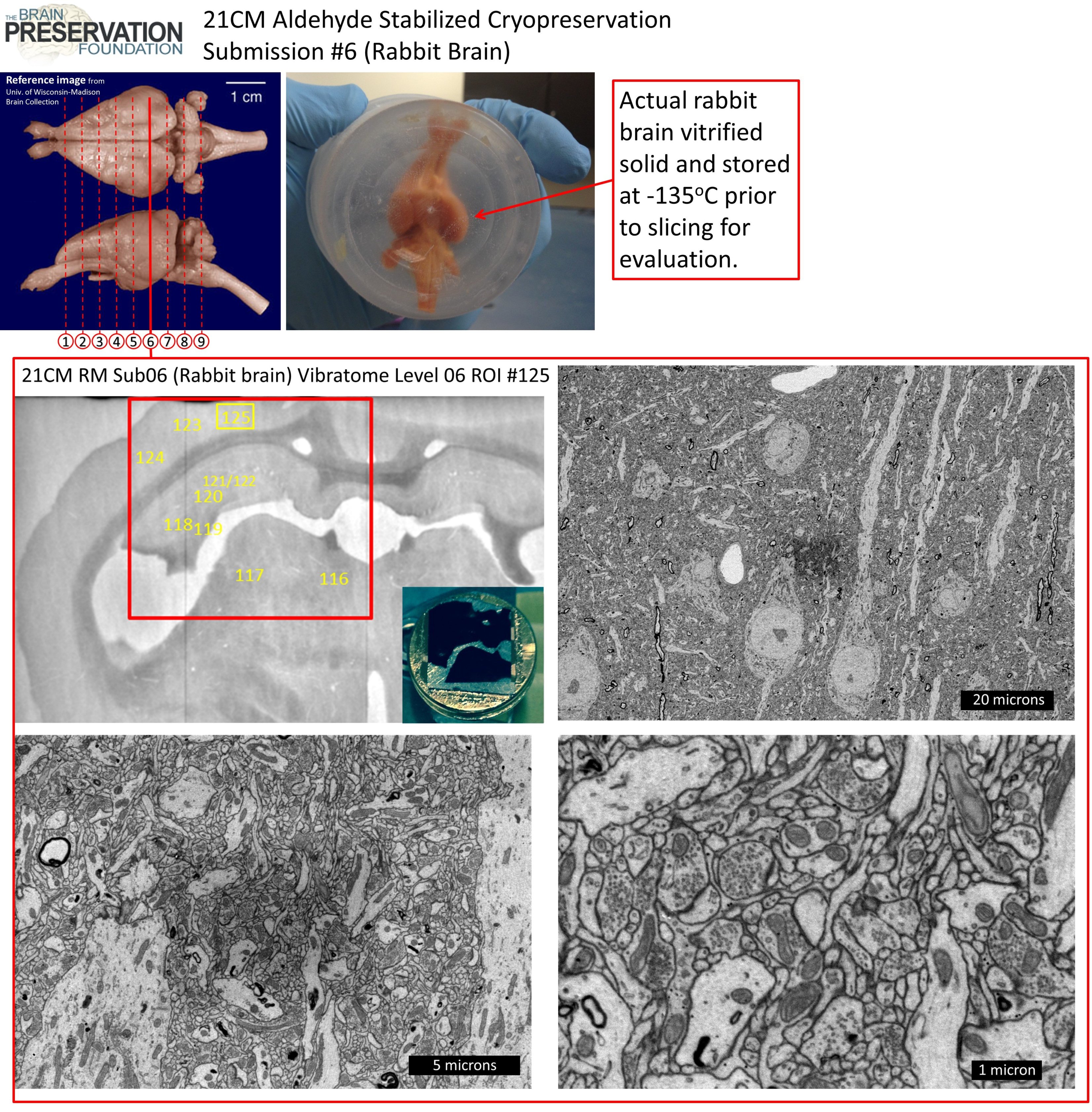
This page contains the detailed electron microscope images taken for evaluation of the Aldehyde-Stabilized Cryopreserved (ASC) rabbit brain (Submission #6) submitted by 21st Century Medicine (21CM) in fulfillment of the requirements for the Brain Preservation Prize. For a summary overview of the ASC procedure itself please refer to our ASC overview page.
Witnessing of preservation procedure
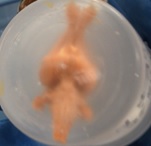
![]() Kenneth Hayworth (KH) (President of the Brain Preservation Foundation (BPF)) and Michael Shermer (member of BPF advisory board) witnessed (on Sept. 25, 2015) the full Aldehyde Stabilized Cryopreservation surgical procedure performed on this rabbit at the laboratories of 21CM under the direction of 21CM lead researcher Robert McIntyre. This included the live rabbit’s carotid arteries being perfused with glutaraldehyde and subsequent perfusion with cryoprotectant agent (CPA). KH witnessed this rabbit brain being put in -135oC storage, removal from storage the following day (verifying that it had vitrified solid), and KH witnessed all subsequent tissue processing steps involved in the evaluation process.
Kenneth Hayworth (KH) (President of the Brain Preservation Foundation (BPF)) and Michael Shermer (member of BPF advisory board) witnessed (on Sept. 25, 2015) the full Aldehyde Stabilized Cryopreservation surgical procedure performed on this rabbit at the laboratories of 21CM under the direction of 21CM lead researcher Robert McIntyre. This included the live rabbit’s carotid arteries being perfused with glutaraldehyde and subsequent perfusion with cryoprotectant agent (CPA). KH witnessed this rabbit brain being put in -135oC storage, removal from storage the following day (verifying that it had vitrified solid), and KH witnessed all subsequent tissue processing steps involved in the evaluation process.
Peer-reviewed publication of procedure
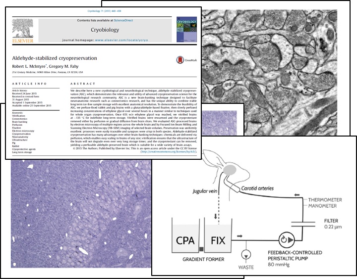 All details of the preservation procedure performed on this rabbit brain have been published in the journal Cryobiology by Robert McIntyre and Greg Fahy in their 2015 paper “Aldehyde-stabilized Cryopreservation”. Here is a link to that open access paper:
http://www.sciencedirect.com/science/article/pii/S001122401500245X
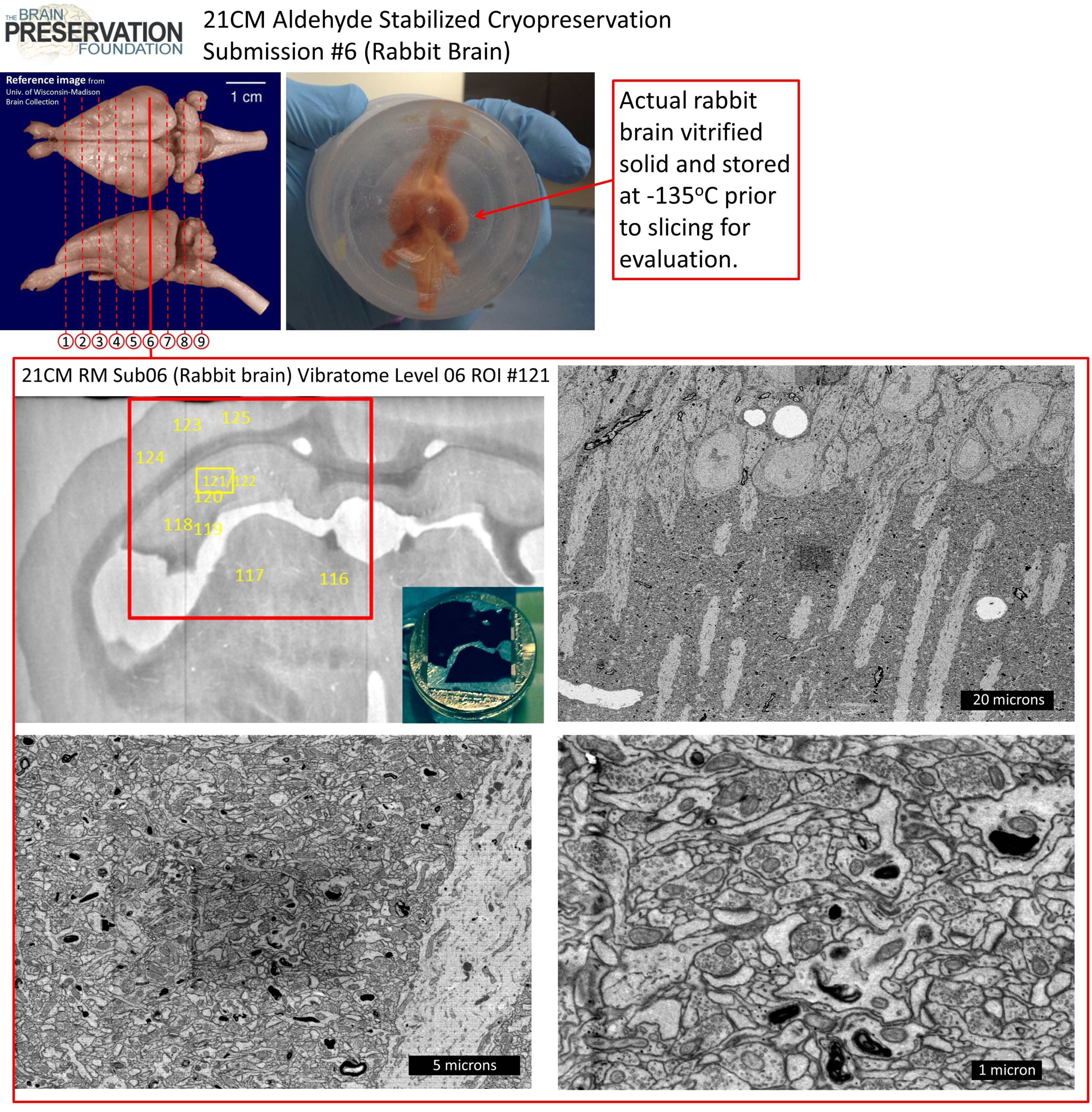
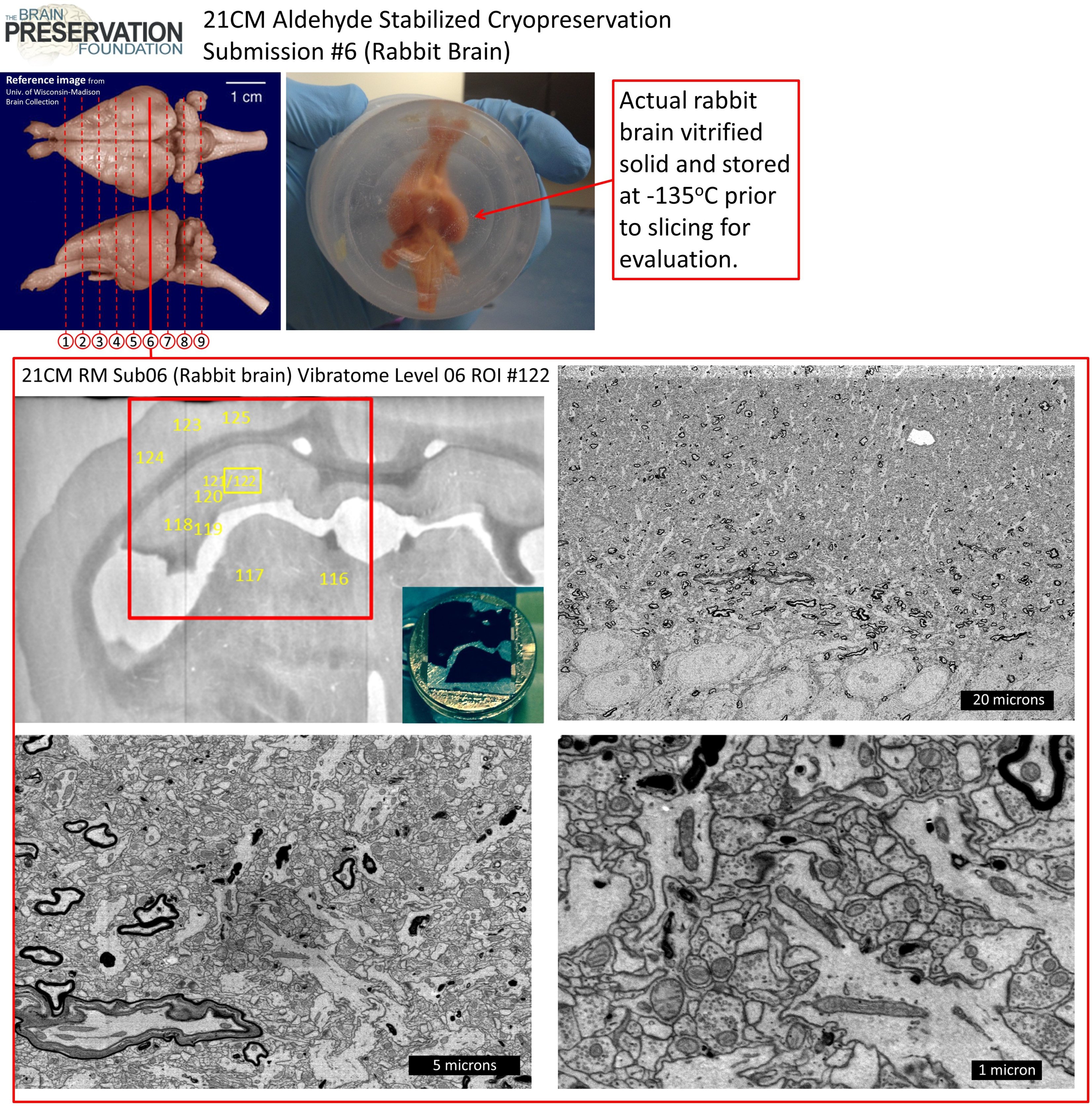
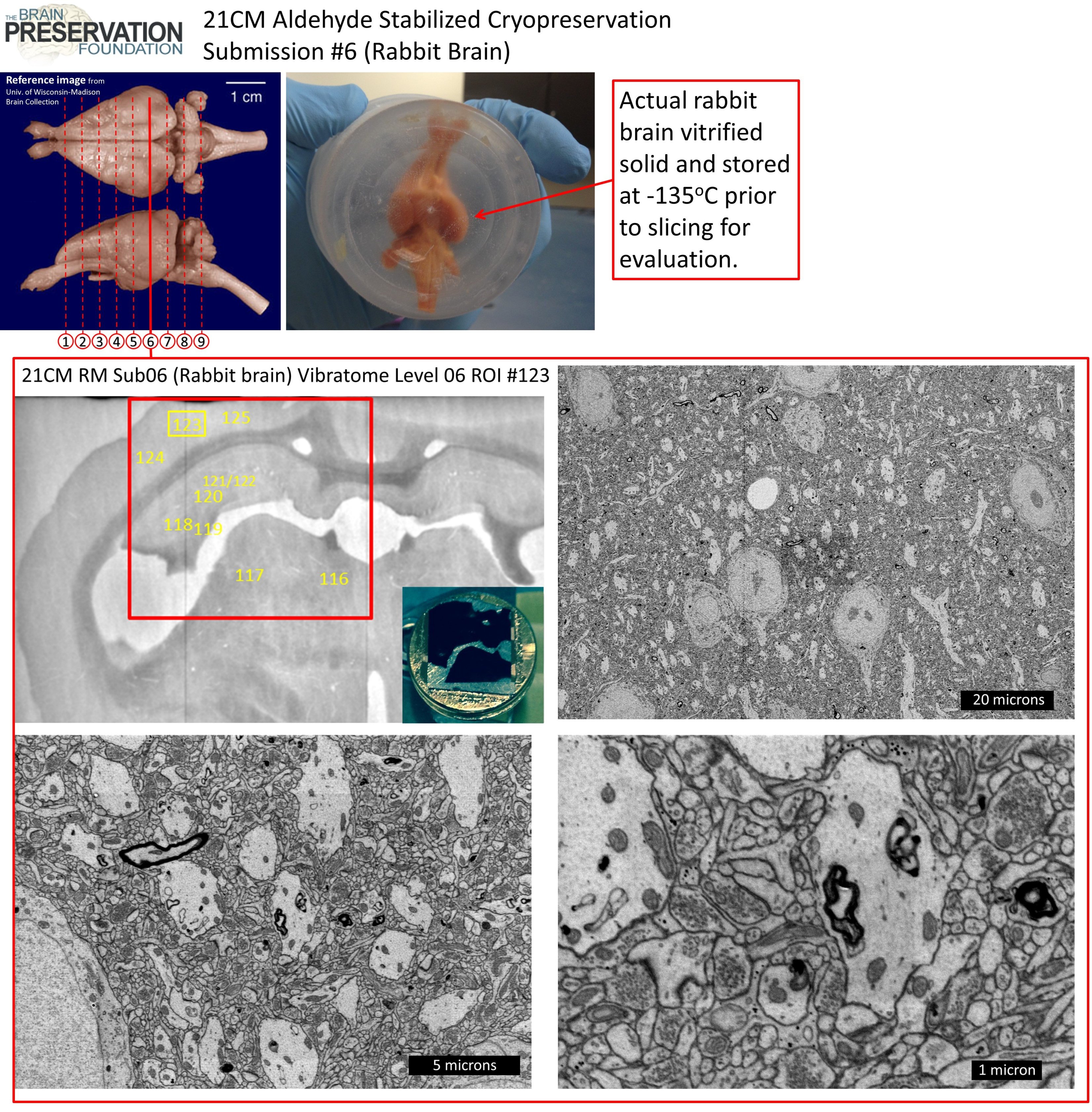
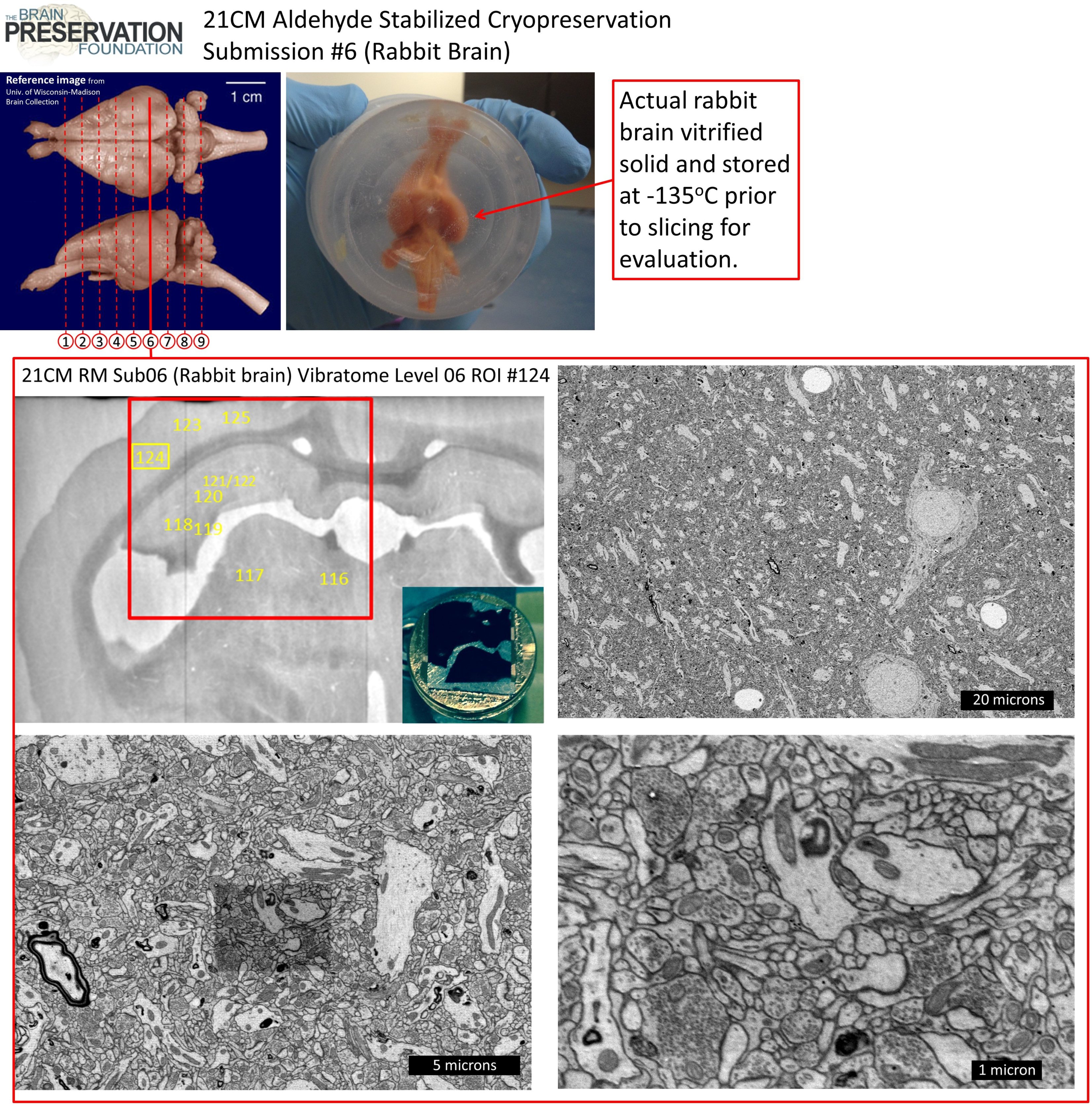
This paper also describes the methods used to vibratome section the rabbit brain into coronal sections, heavy metal stain those sections, and process them for electron microscopy -the same methods used to obtain the images seen in this evaluation page.
Link to download all evaluation FIB-SEM movies and Electron Micrographs
In order to verify that the brain ultrastructure of this Aldehyde Stabilized Cryopreserved rabbit brain was in fact well preserved required extensive electron microscope imaging from widely spaced regions of the rabbit’s brain. Individual links to many of these movies and images are provided on this web page.
We have also put all of the 3D datasets and images into a Google Drive link: CLICK HERE FOR GOOGLE DRIVE LINK TO DATASETS AND IMAGES
Movies of intact rabbit brain
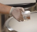 A movie (taken by KH) of the rabbit brain floating in fixative+CPA after being removed from skull, being put into a -135oC freezer storage unit.
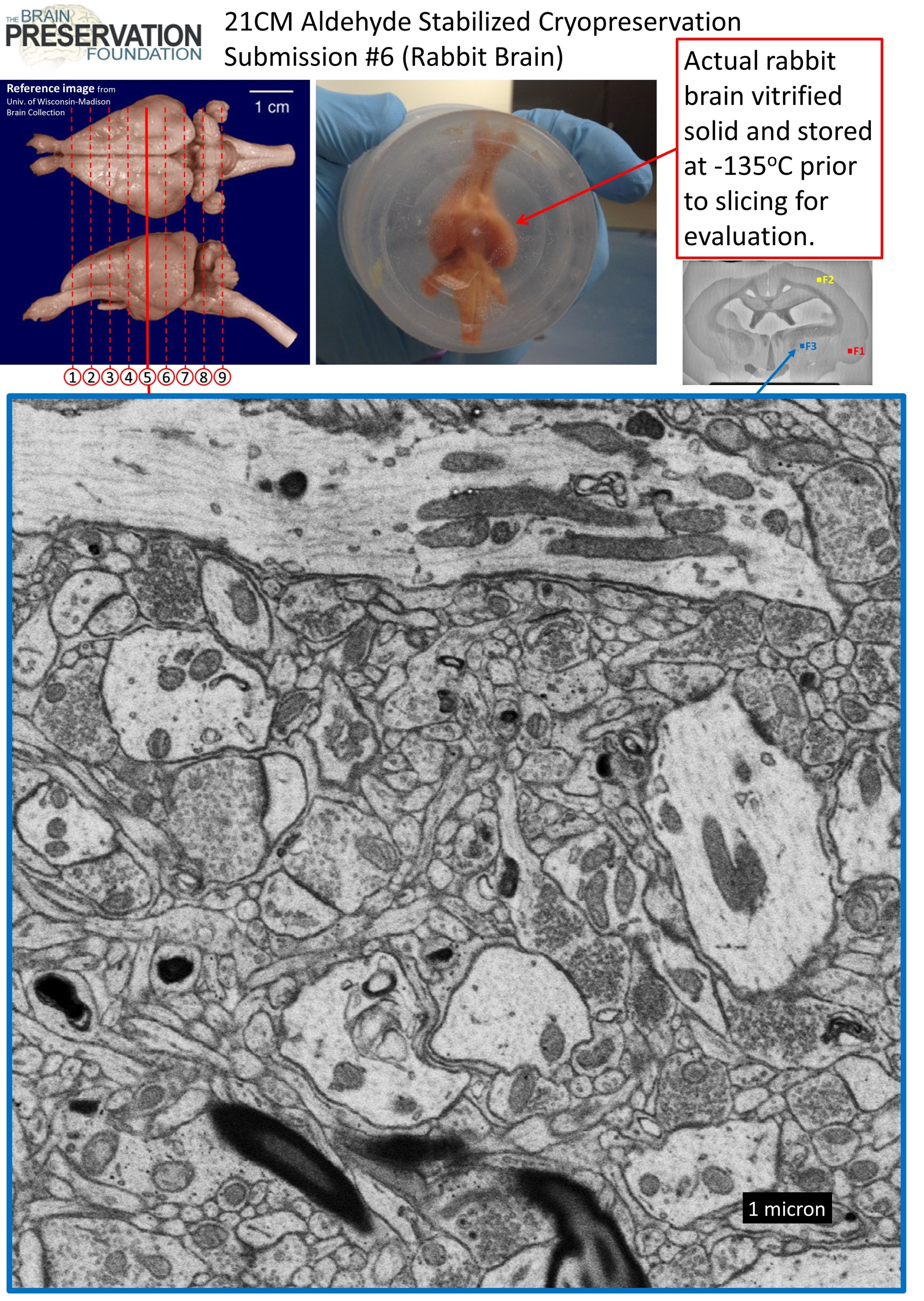
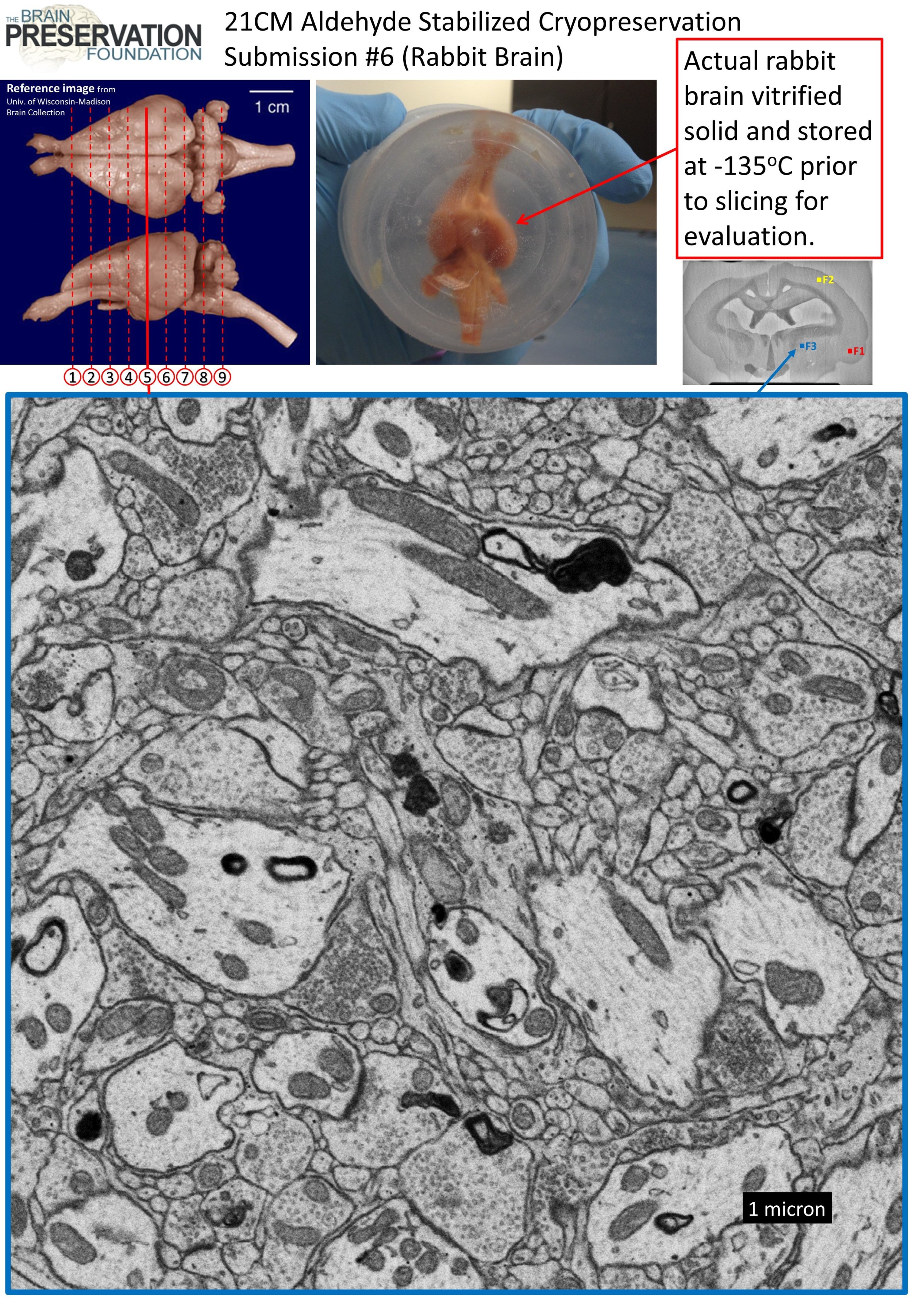
 A movie (taken by KH) of that same rabbit brain being taken out of the -135oC freezer unit after overnight storage. It is clearly visible that the brain has vitrified solid at this temperature.
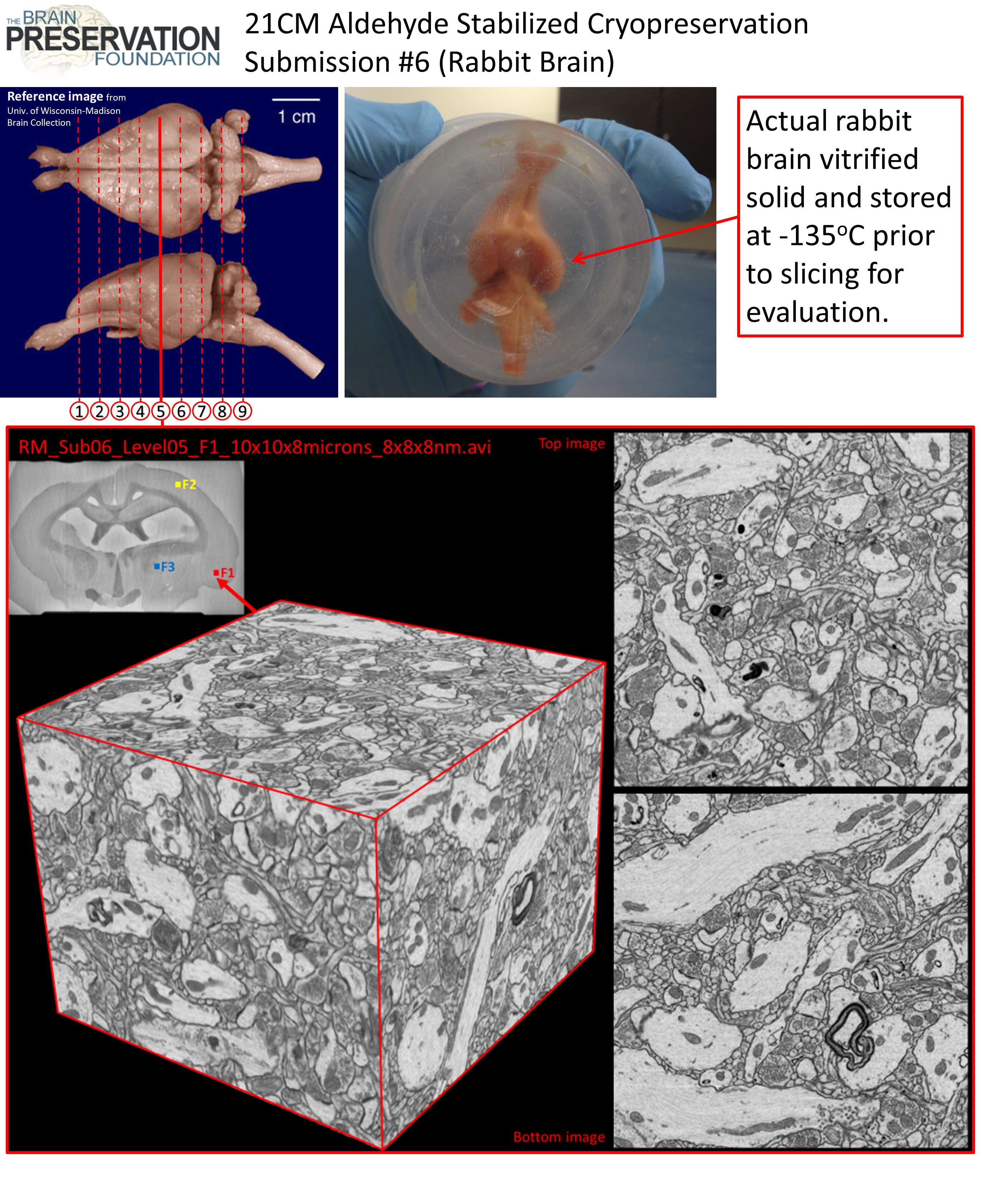
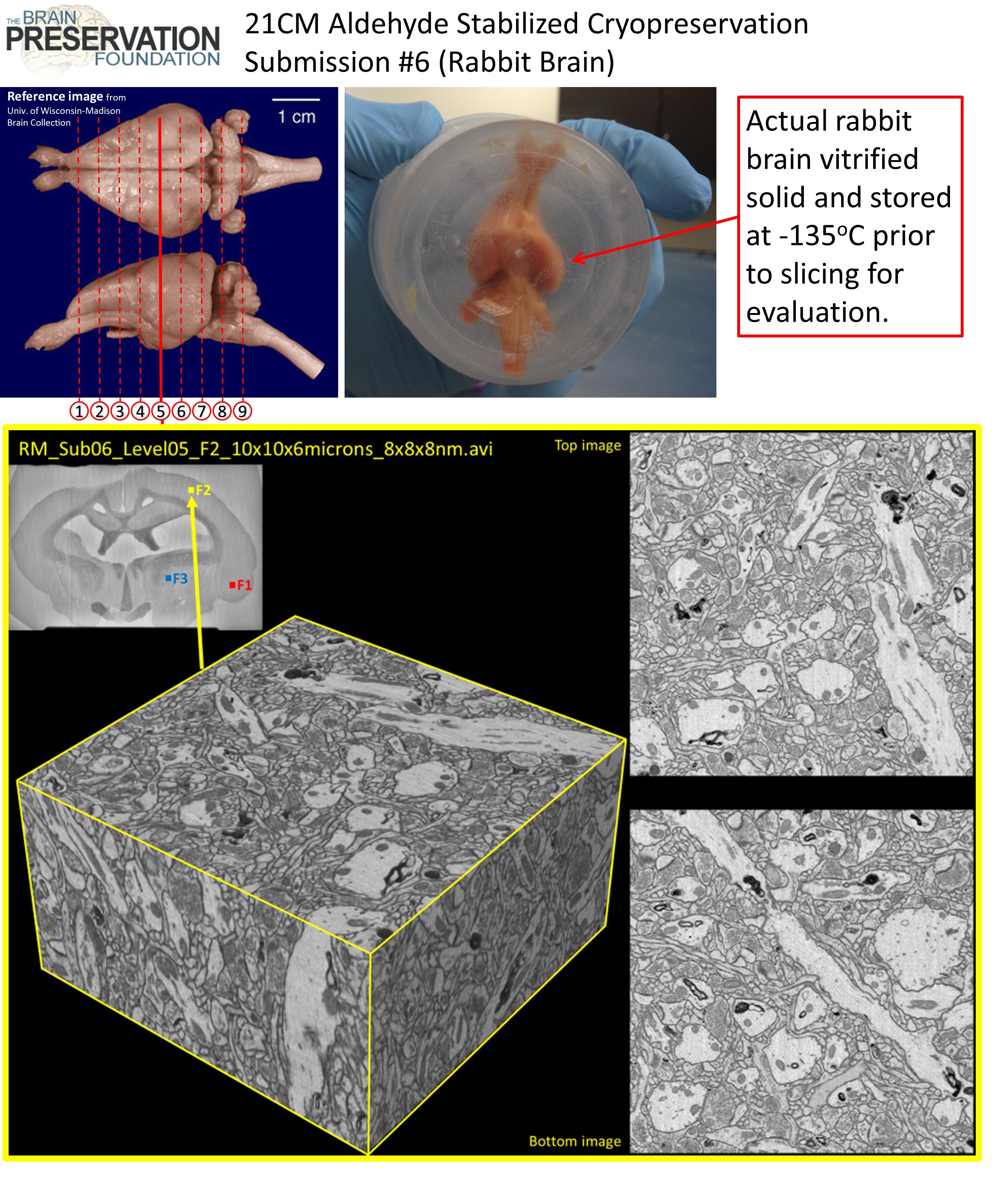
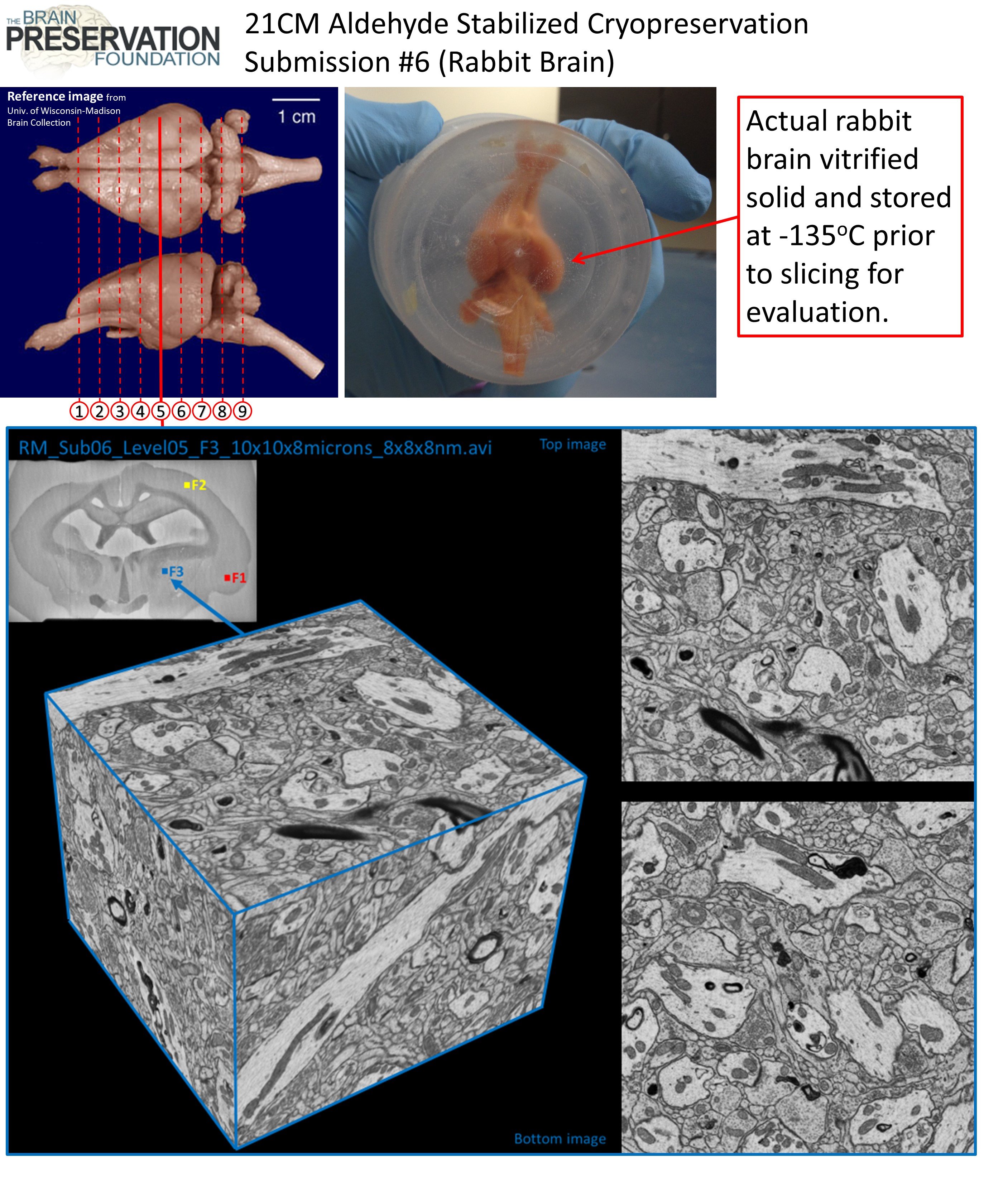
Focused Ion Beam Scanning Electron Microscopy (FIB-SEM) Evaluation Volumes
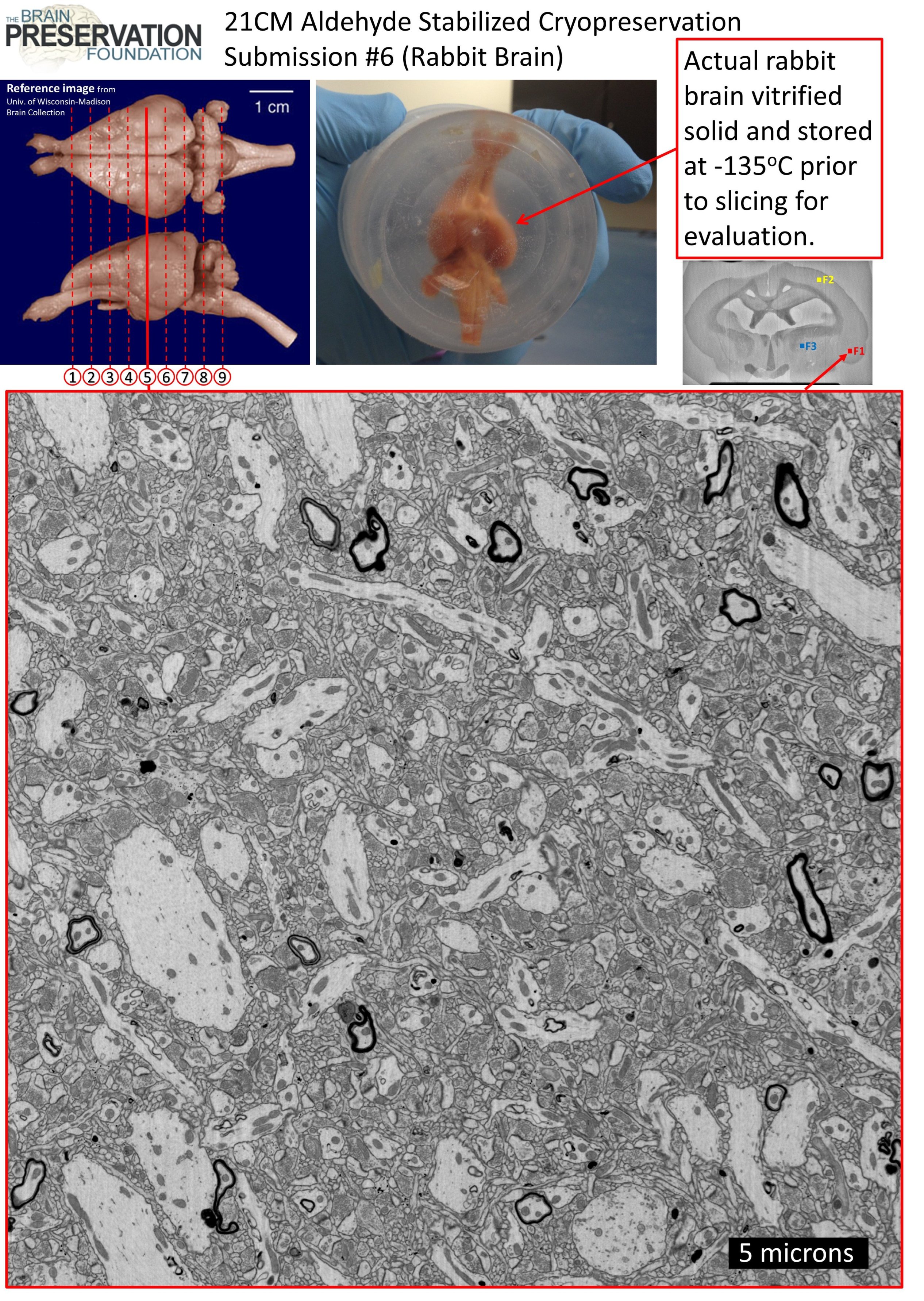
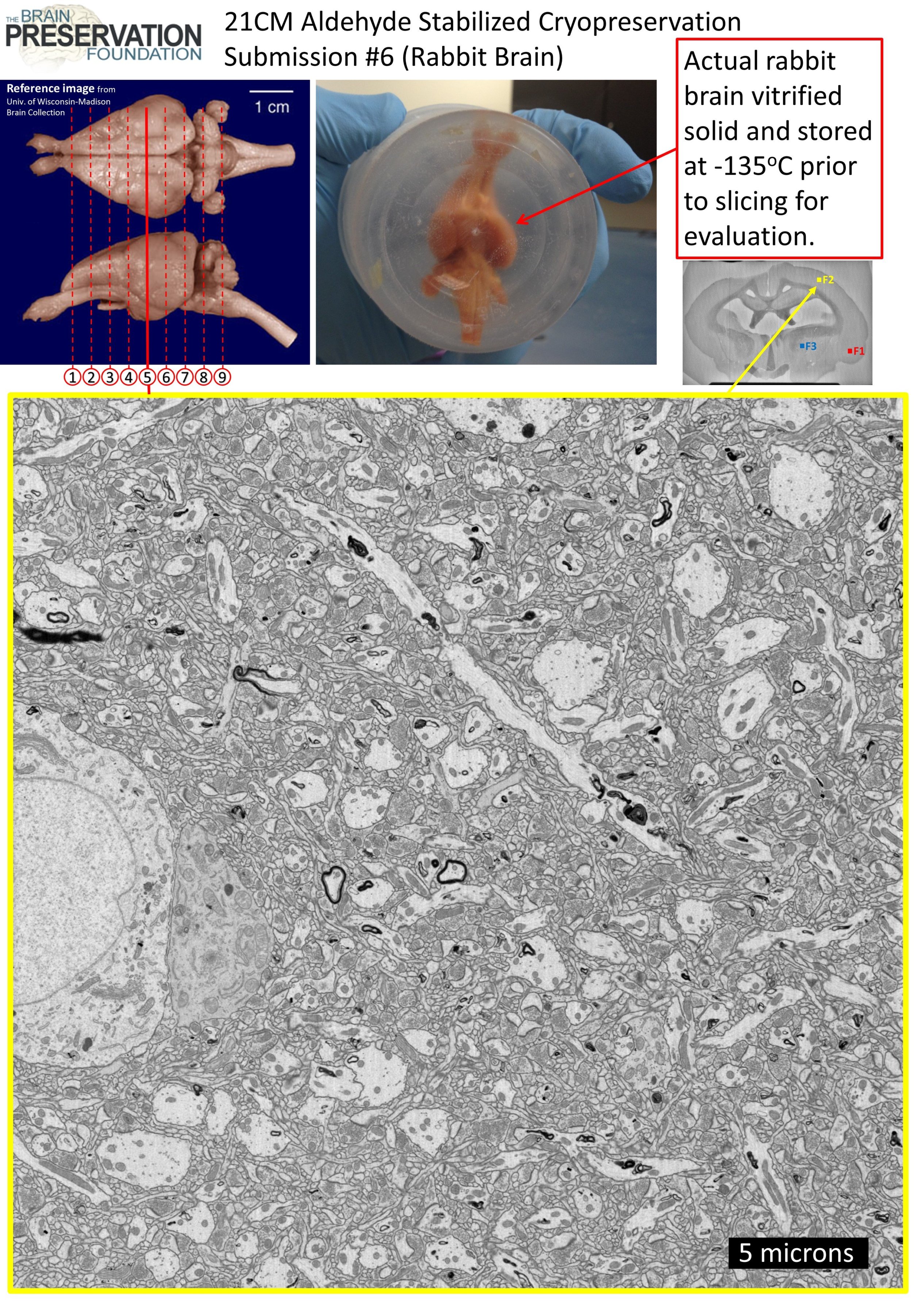
As part of the prize requirements we needed to evaluate whether the 3D synaptic connectivity of the rabbit brain was well preserved. To accomplish this (KH) acquired three FIB-SEM volumes from widely separated parts of a coronal vibratome section midway through the ASC rabbit brain. An overview of the FIB-SEM imaging process can be obtained in (Hayworth et al. 2015).
FIB-SEM volume ‘F1’ cortex:


Link to download movie: https://spaces.hightail.com/space/kYOGB

Link to download movie: https://spaces.hightail.com/space/kYOGB
FIB-SEM volume ‘F2’ cortex:
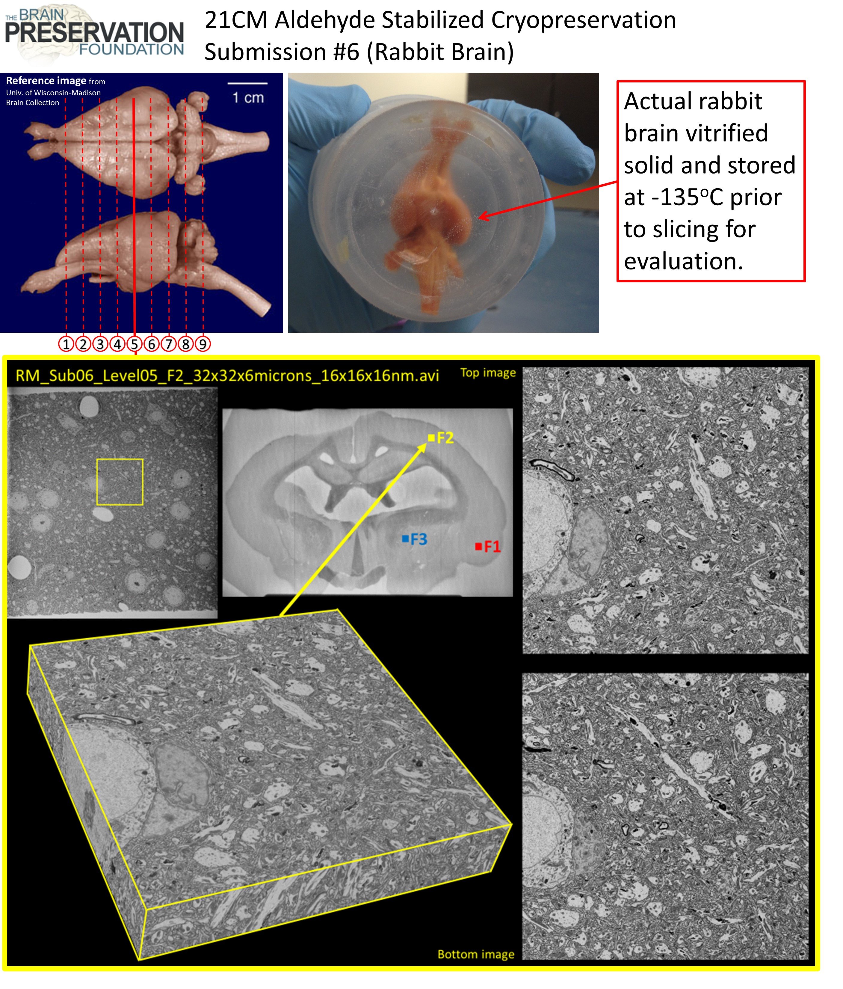

Link to download movie: https://spaces.hightail.com/space/kYOGB
Link to download movie: https://spaces.hightail.com/space/kYOGB
FIB-SEM volume ‘F3’ striatum:
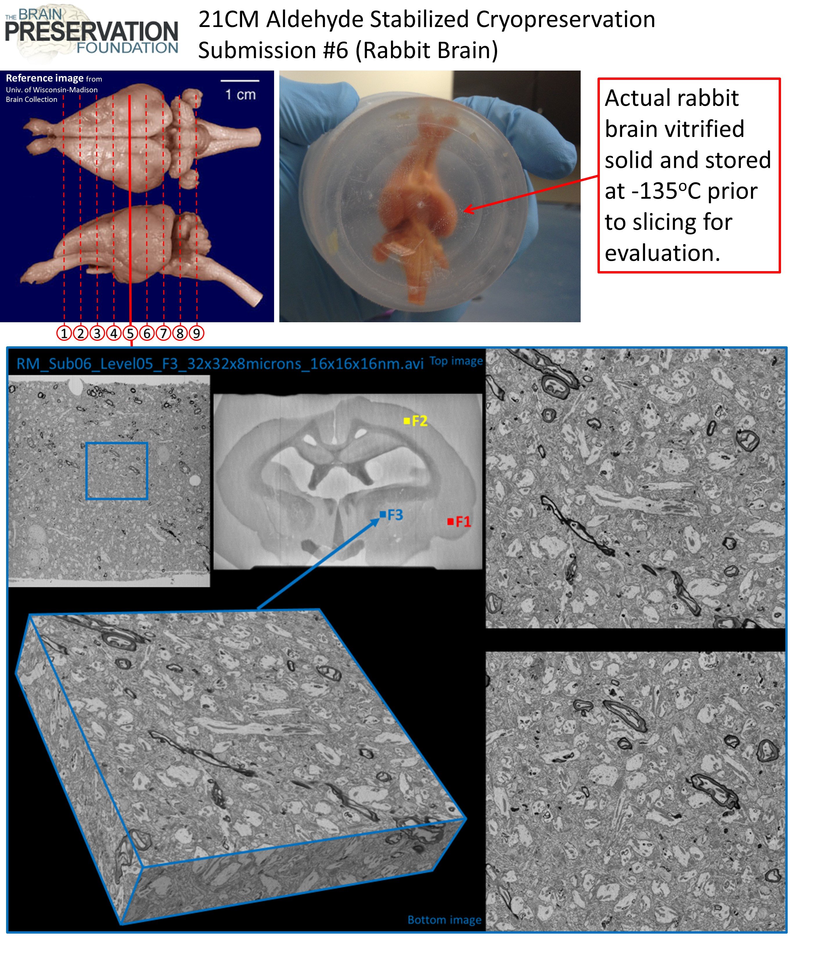
Link to download movie: https://spaces.hightail.com/space/kYOGB
Link to download movie: https://spaces.hightail.com/space/kYOGB
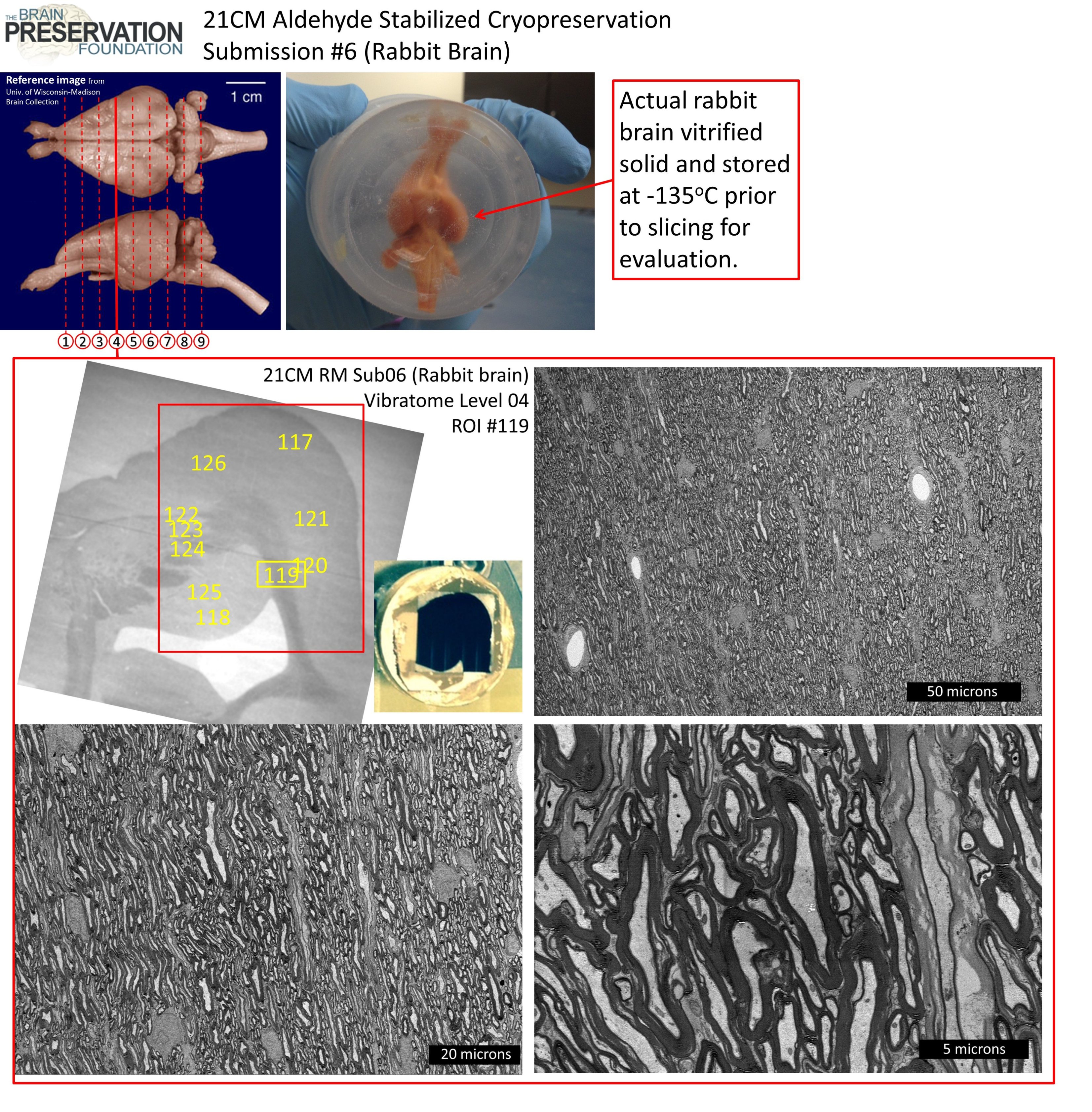
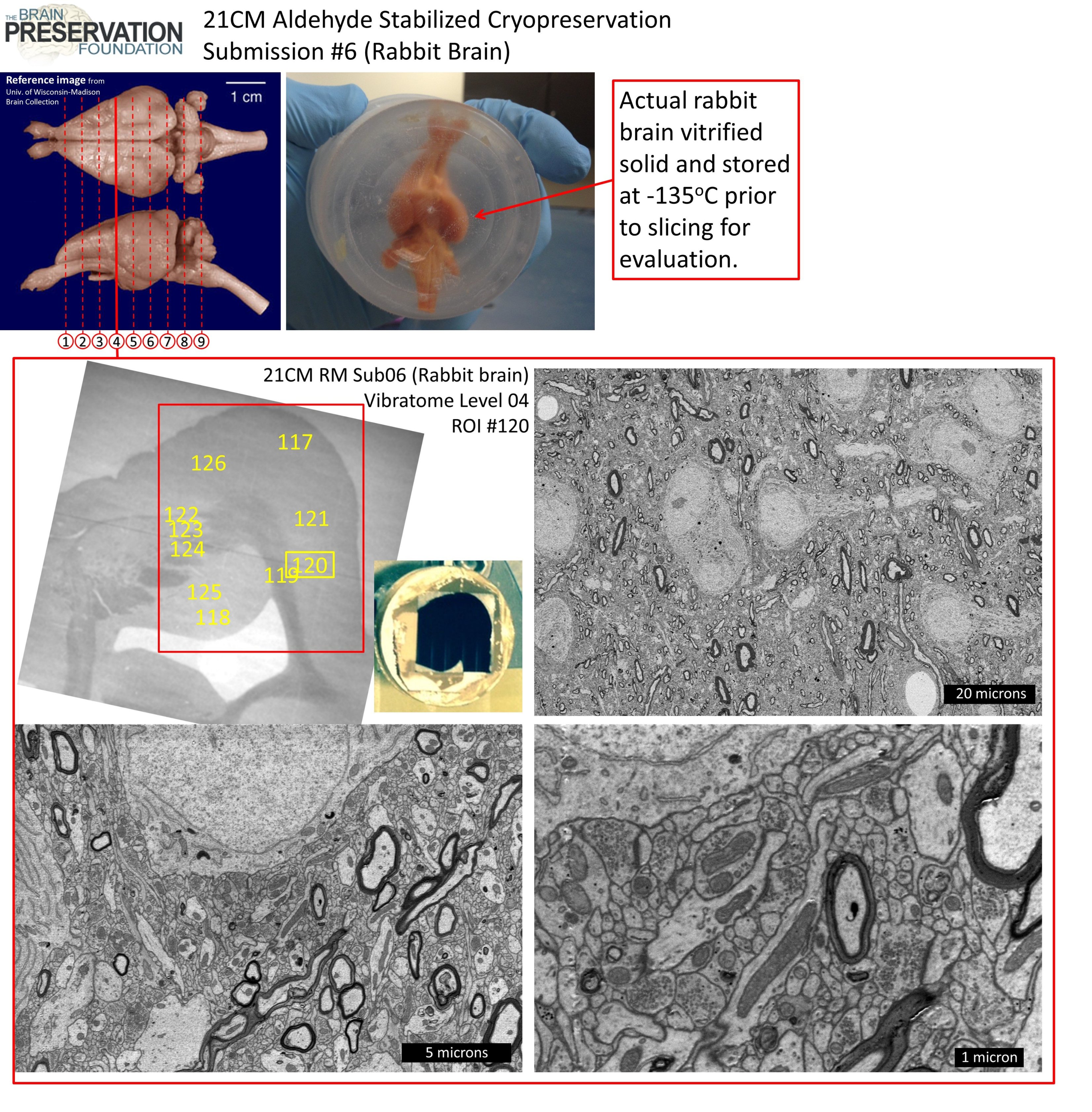
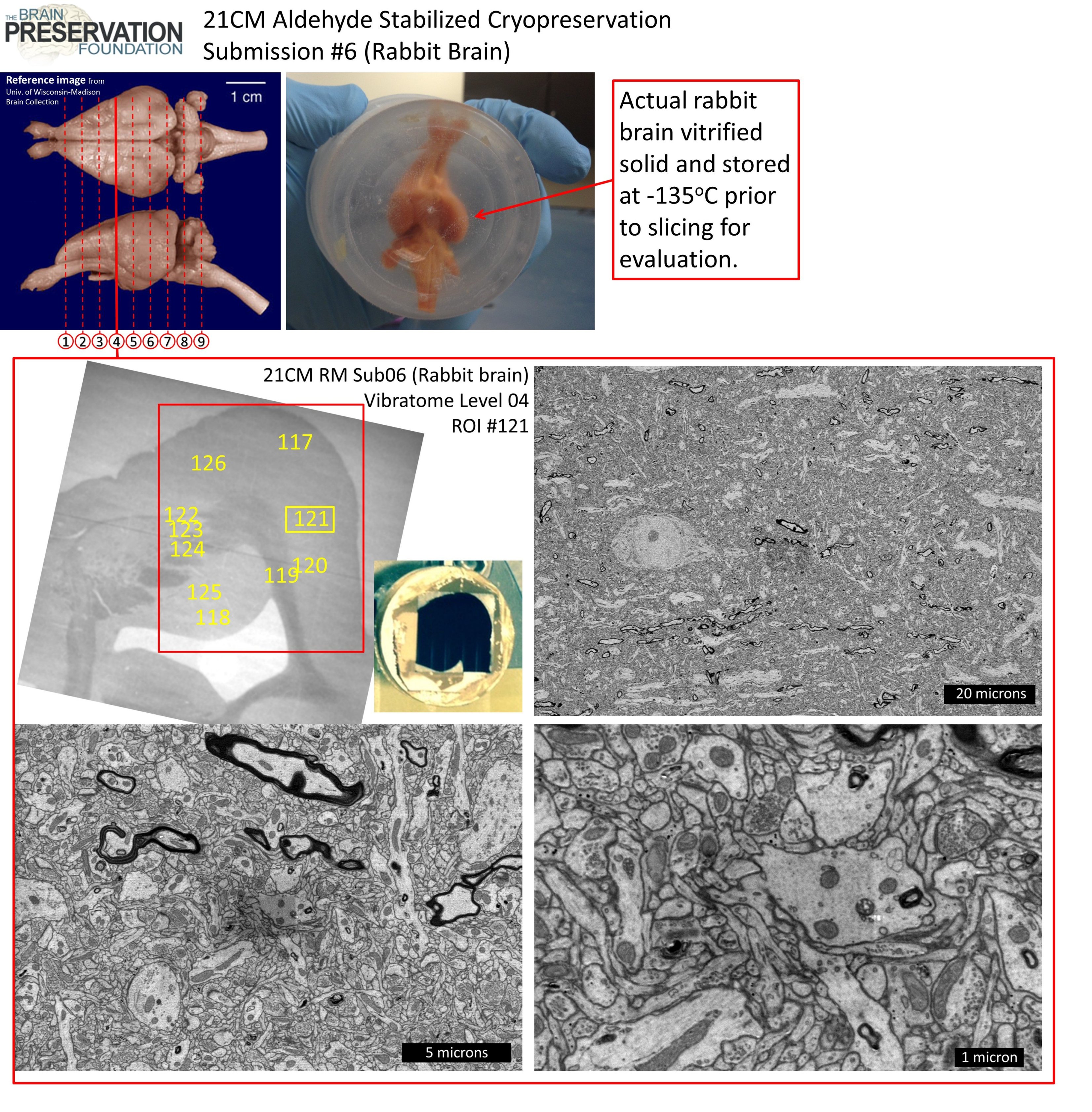
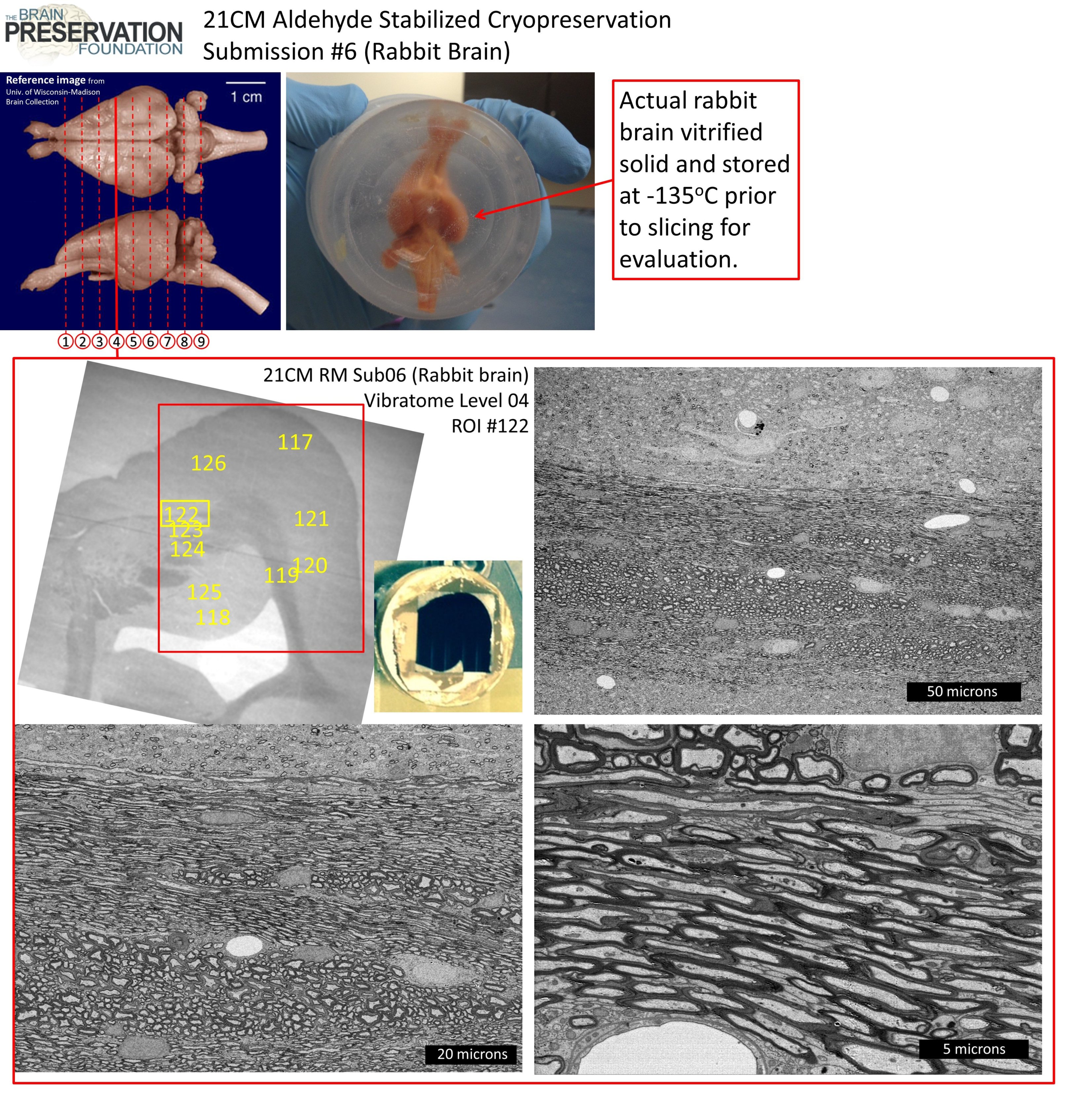
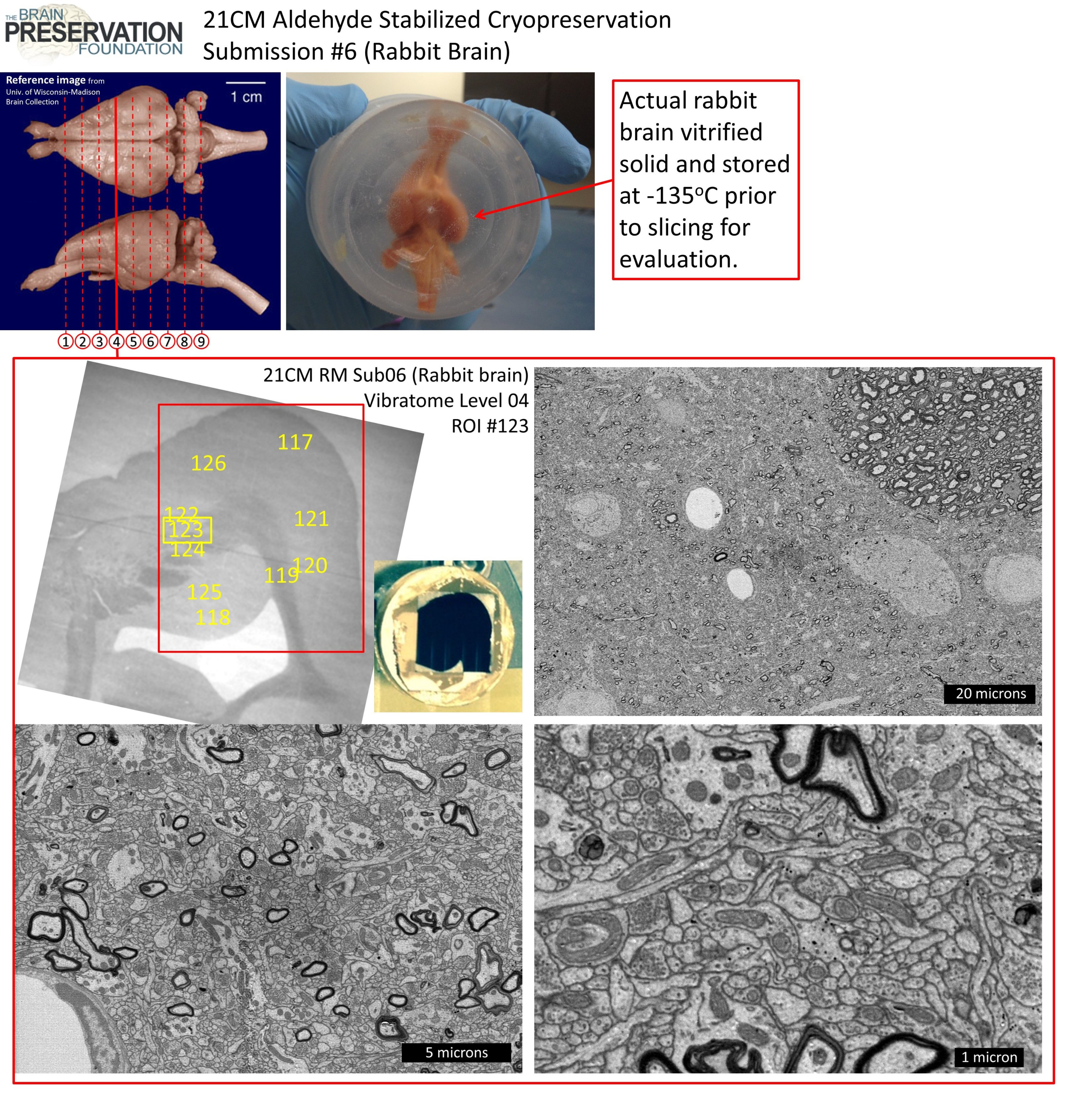
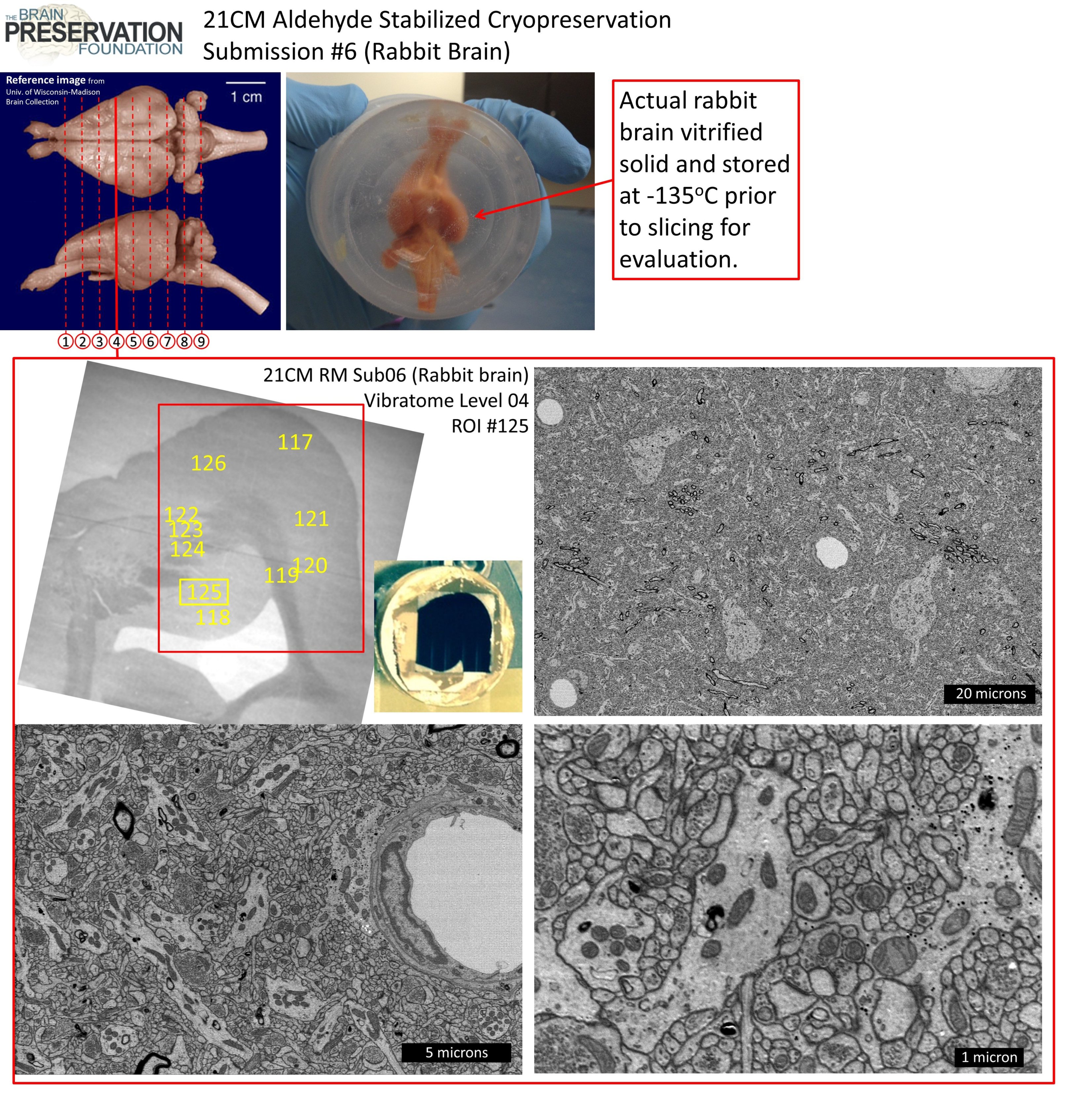
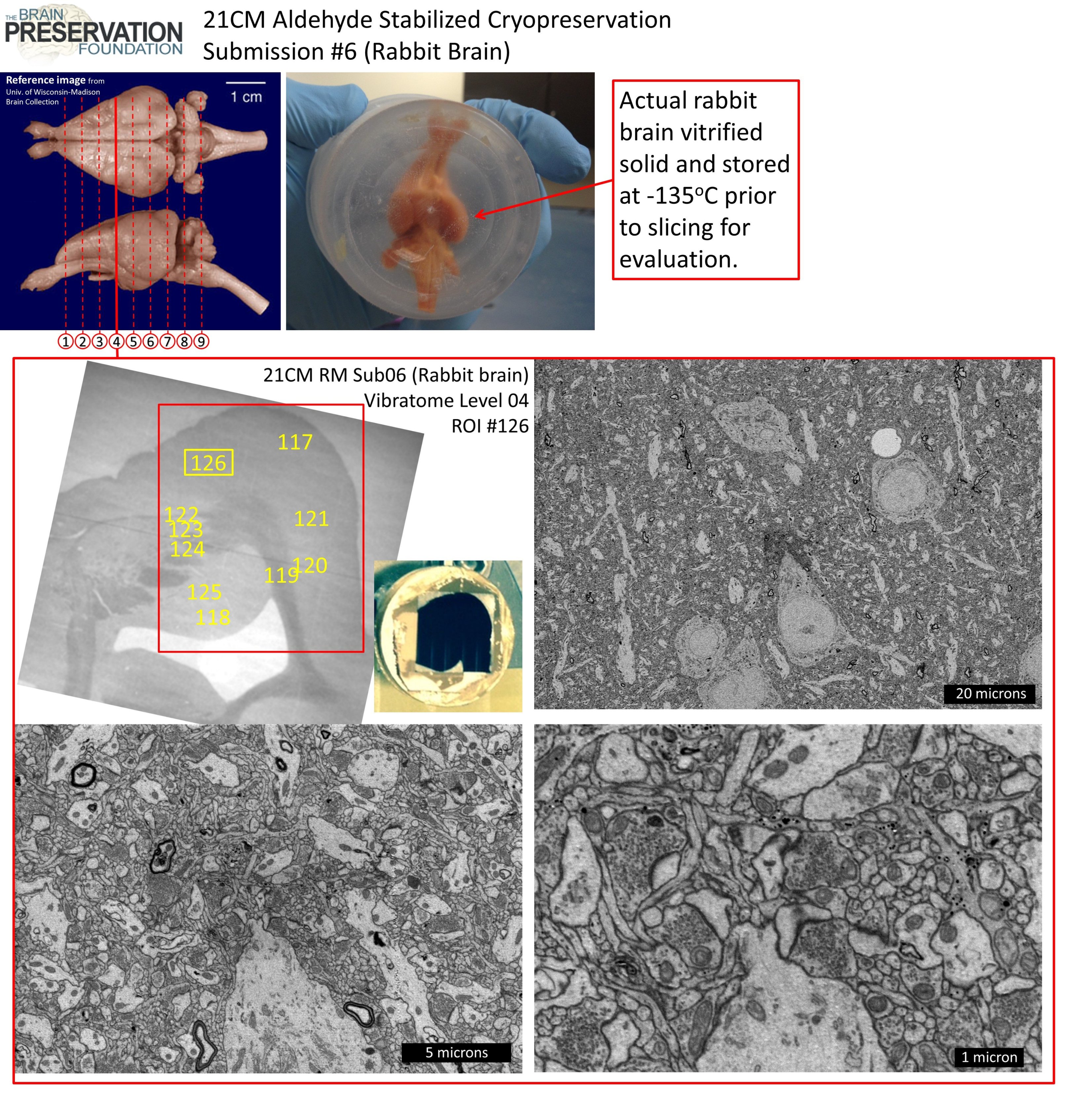
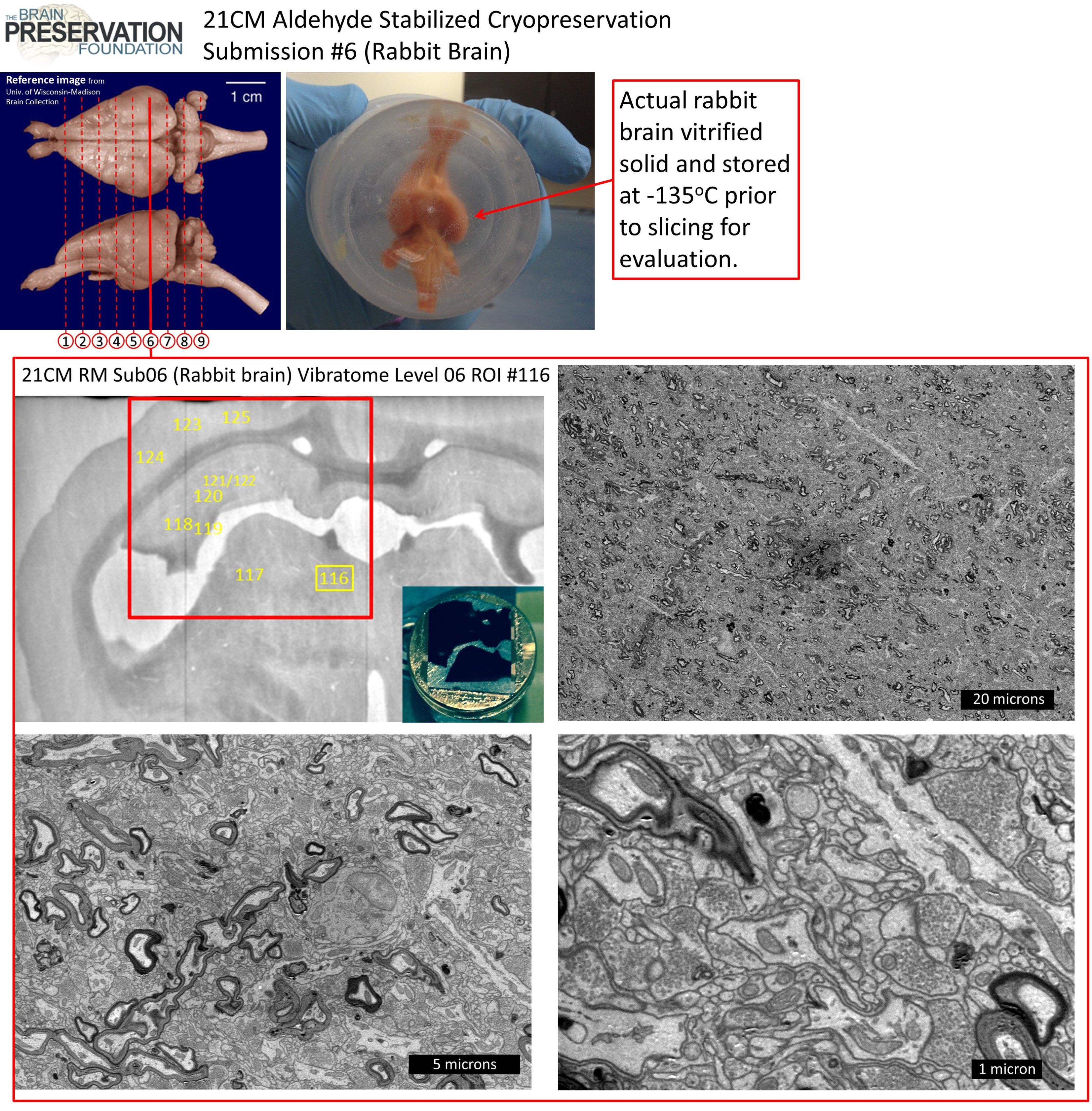
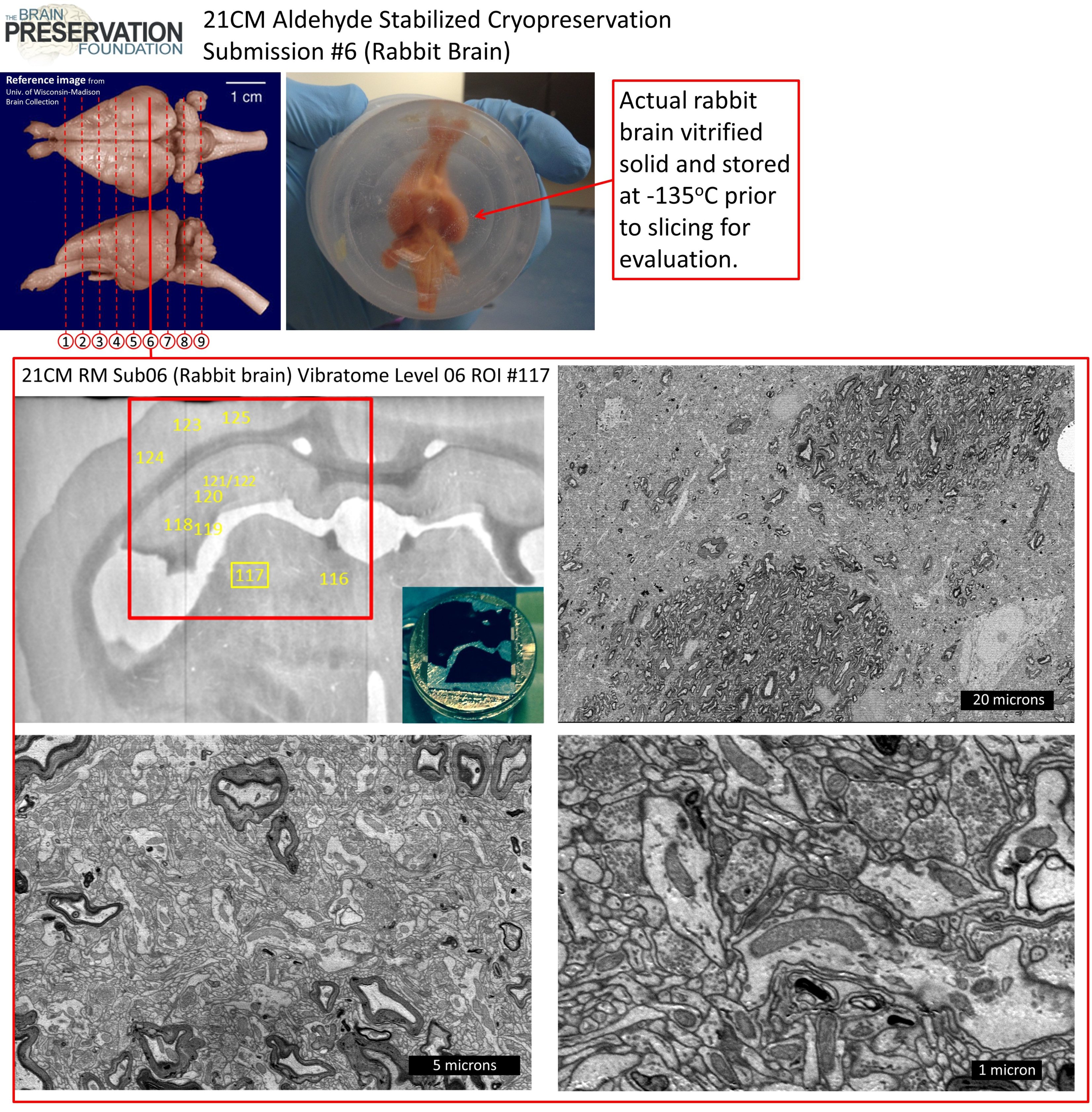
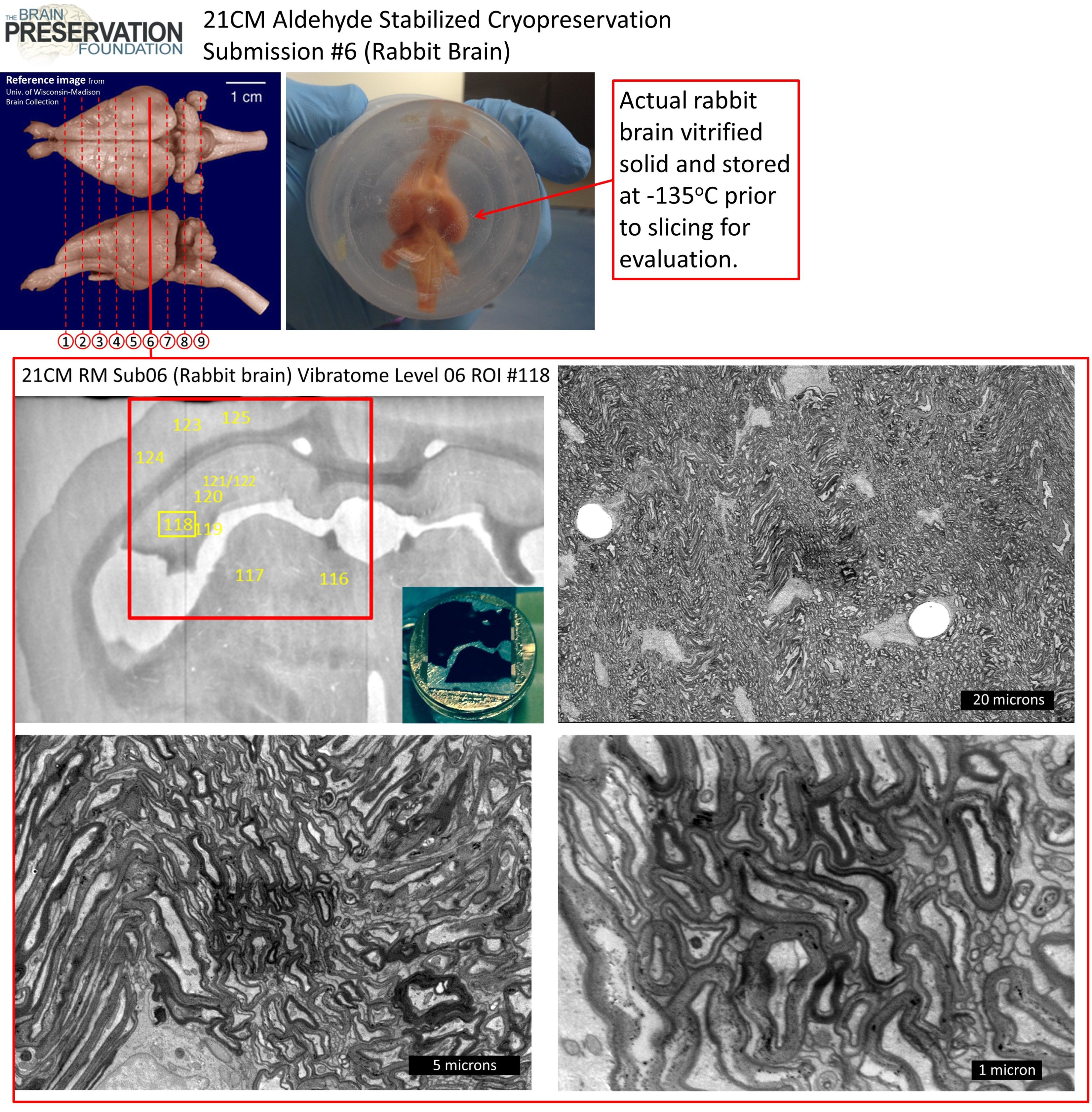
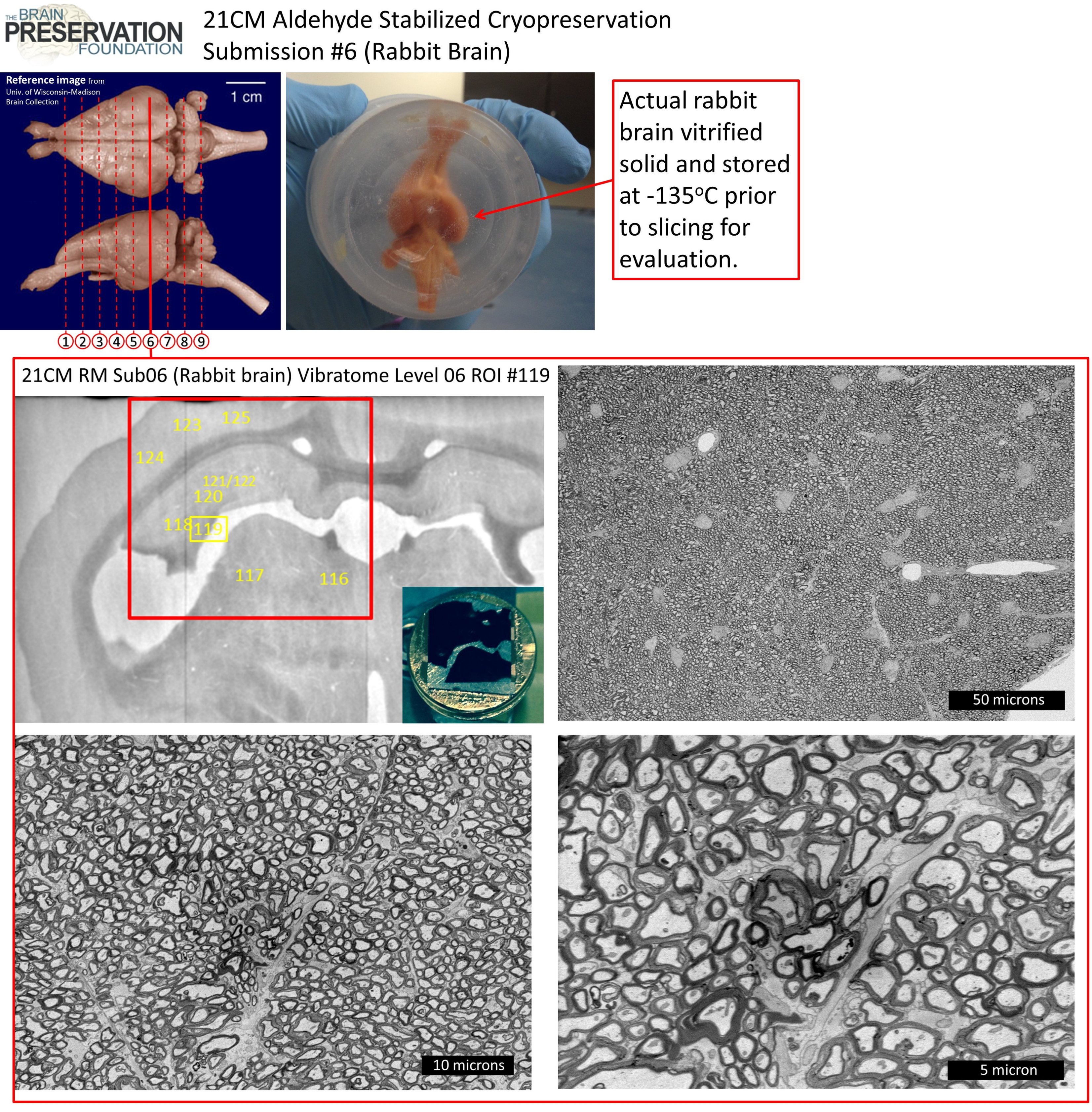
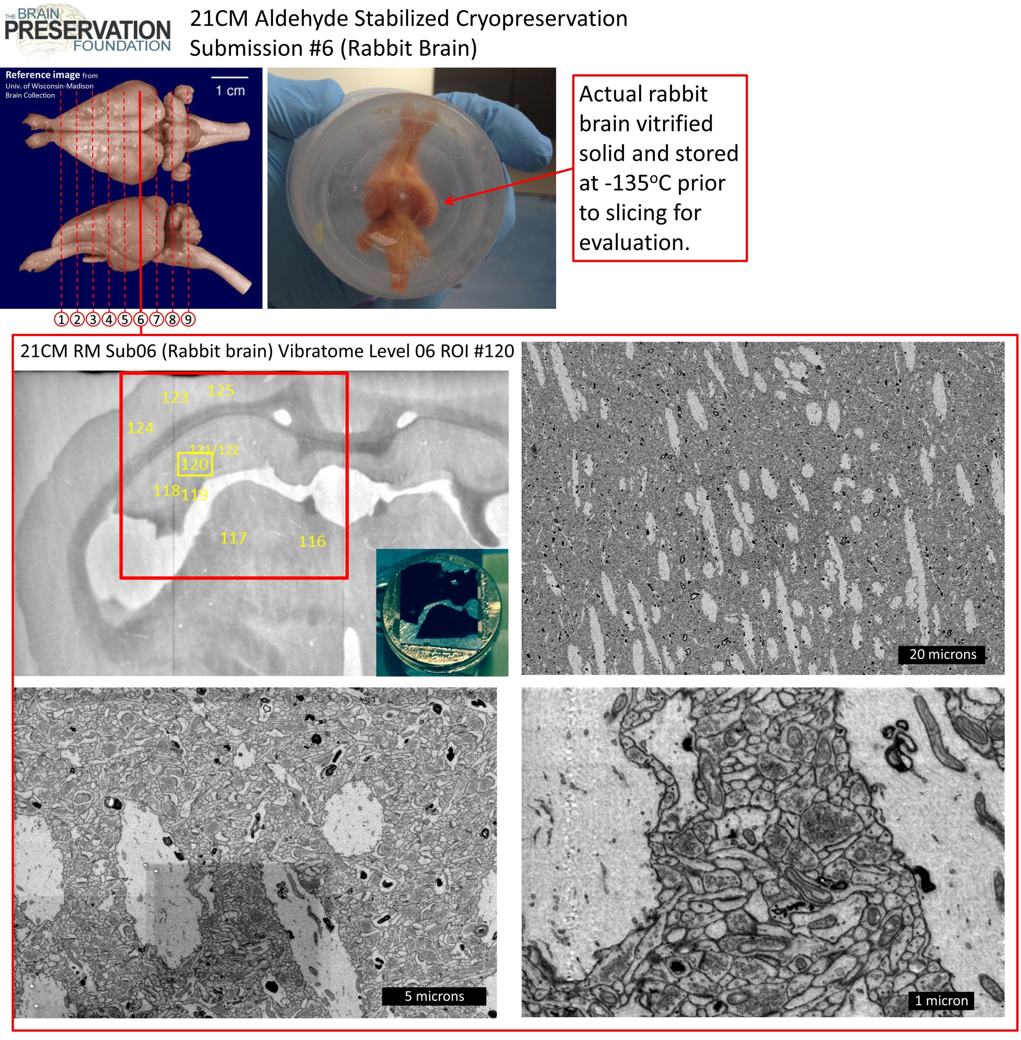
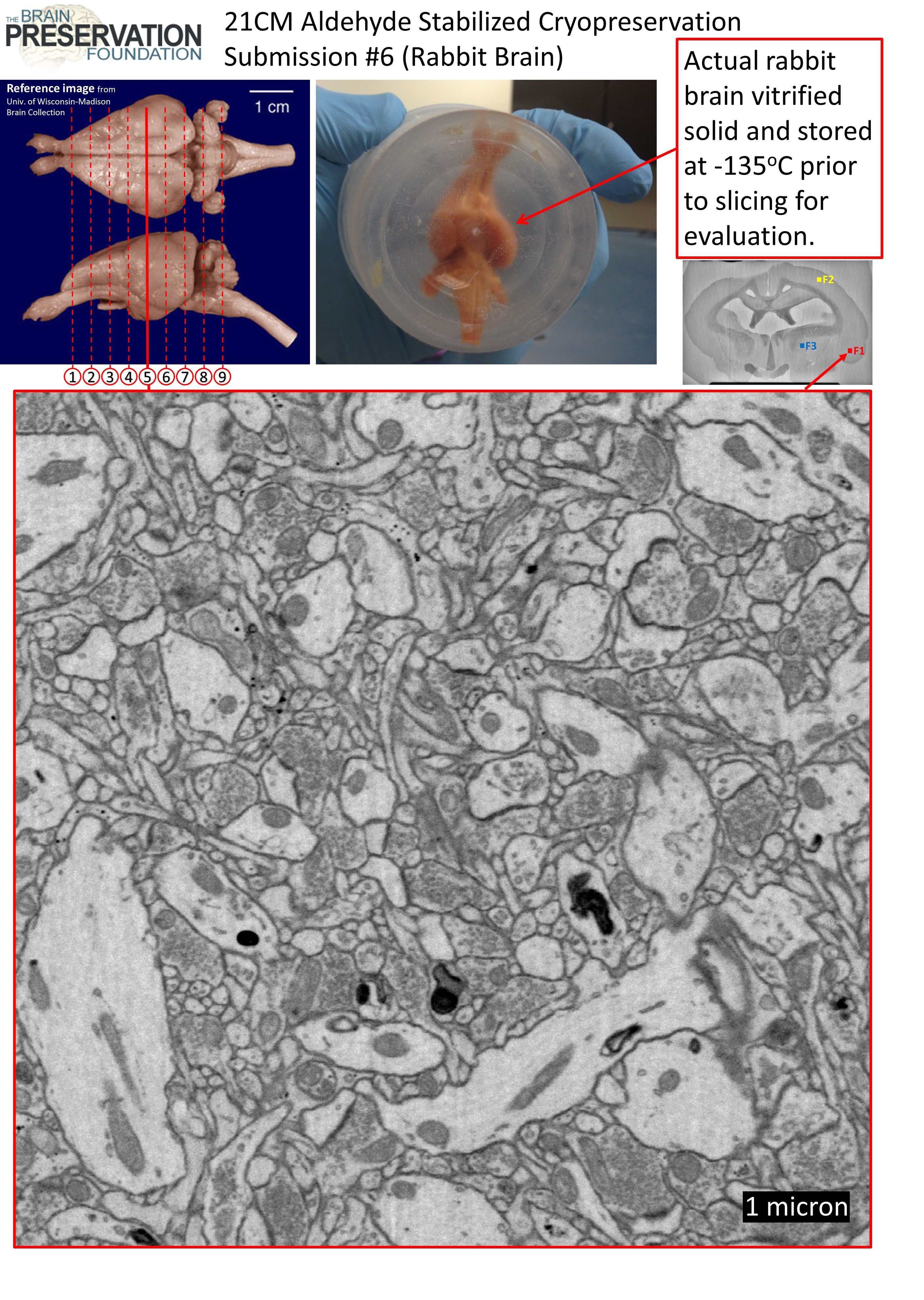
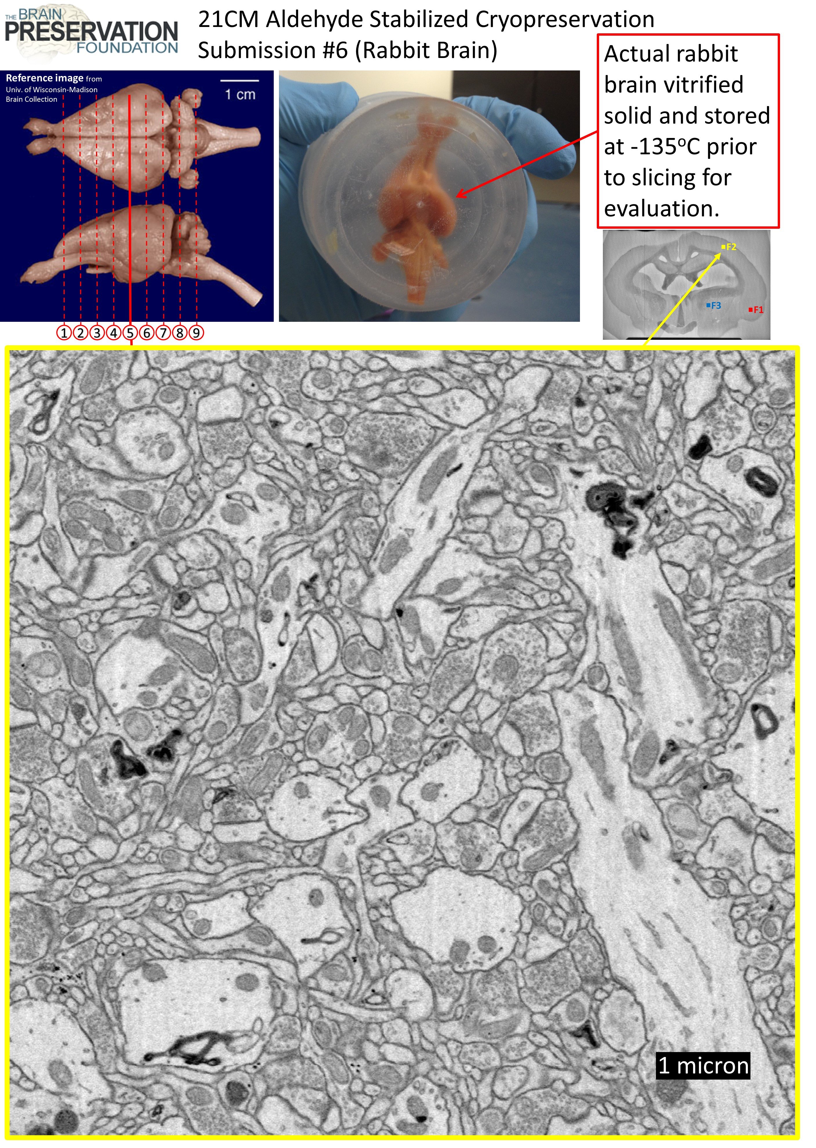
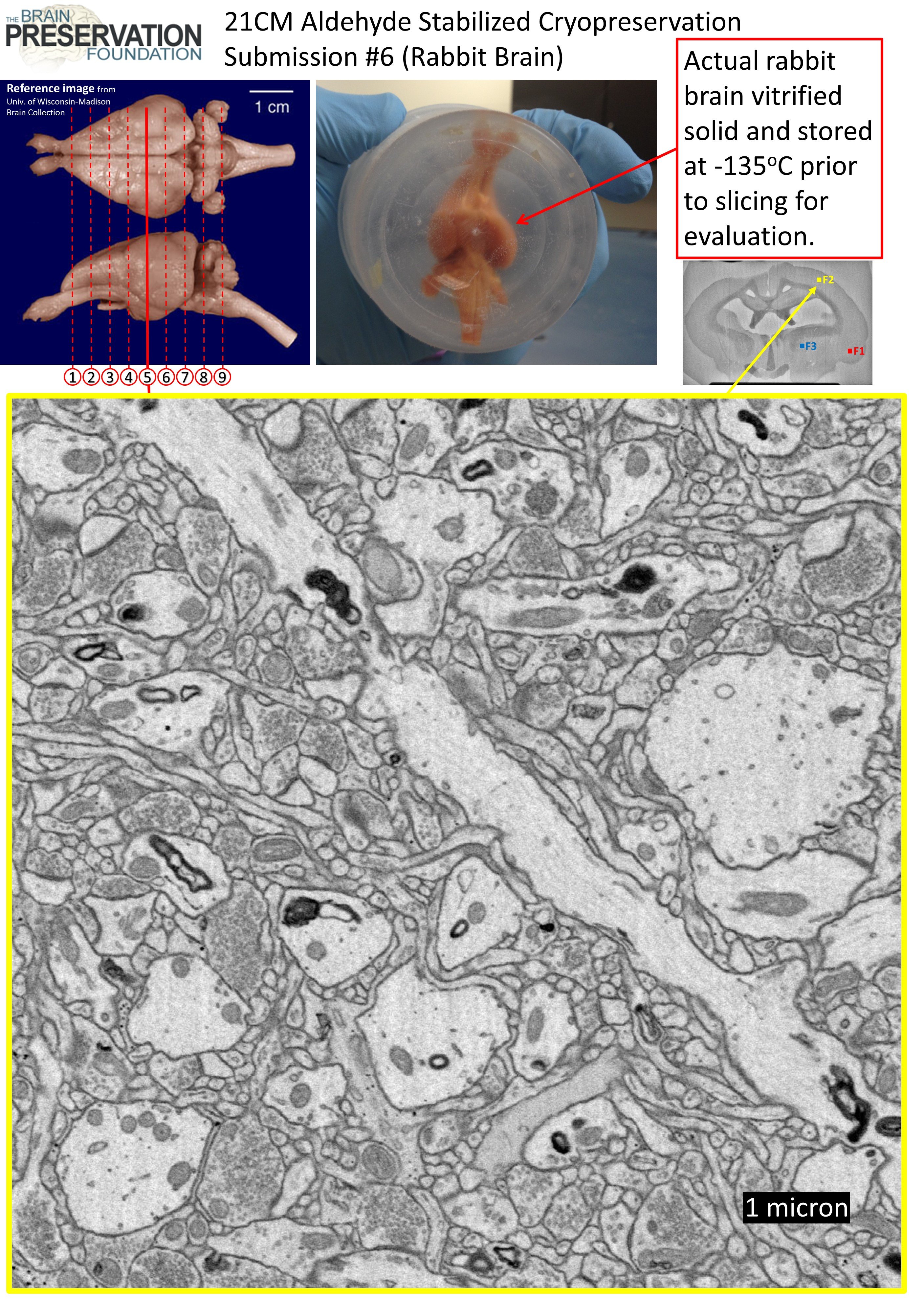
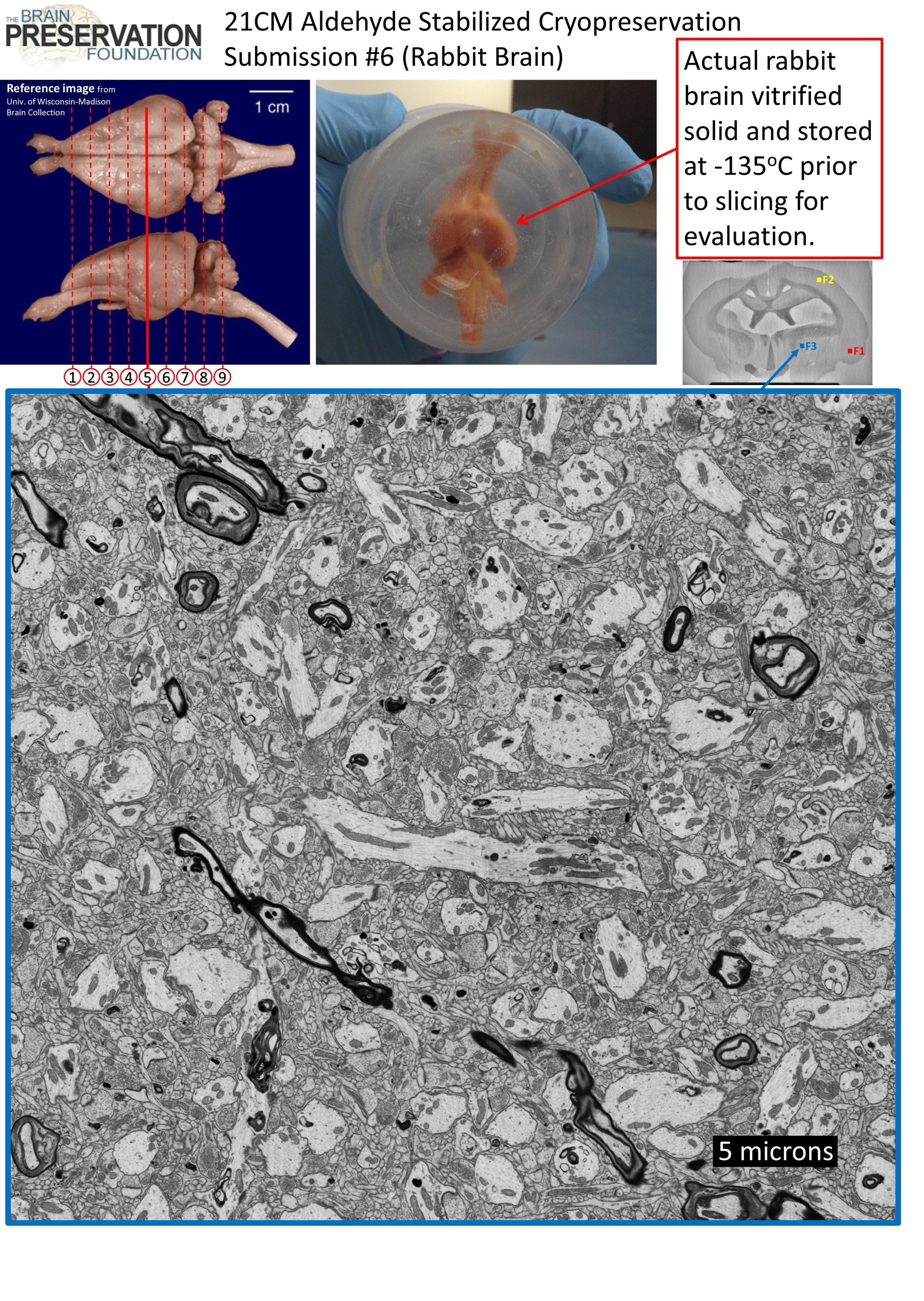
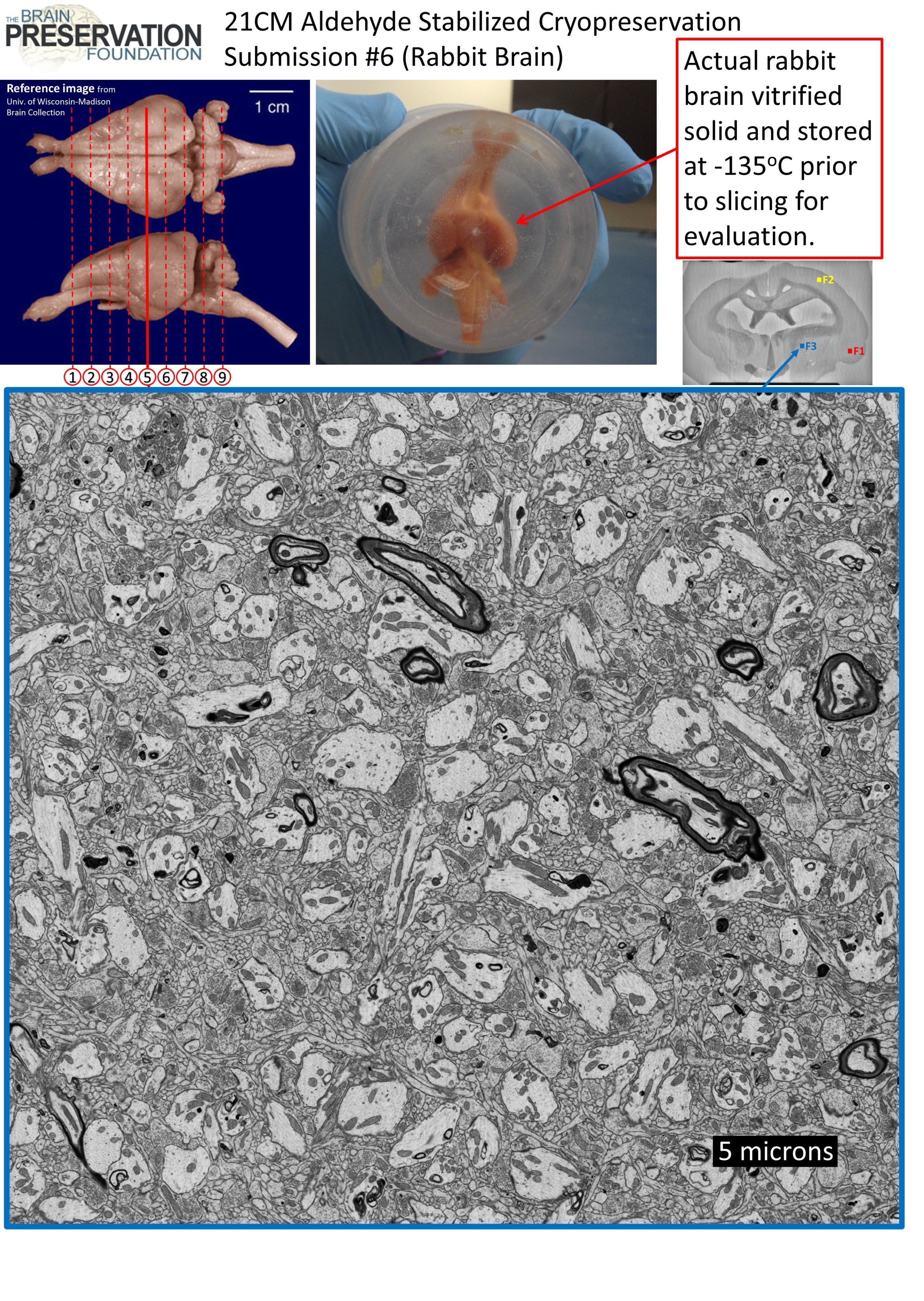
2D SEM survey images
These are a subset of the 2D Scanning Electron Microscope survey images we performed on vibratome slices 4 and 6. These include images of cortex, basal ganglia, hippocampus, and thalamus. Additional survey images are contained in the evaluation zip file: https://spaces.hightail.com/space/QOGUe