
Simulating motion detection in the Drosophila visual system with connectome data
A new manuscript from Gornet et al. describes their work using a serial electron micrograph-derived Drosophila connectome to simulate motion-detection in neurons in the T4 area of the optic lobe:
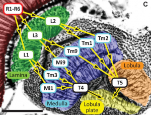
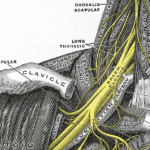
Horizontal section of the Drosophila optic lobe; from https://doi.org/10.7554/eLife.24394.002
They used the simulation tool NEURON, which is a wonderful and widely-used tool for predicting the electrical output from well-defined sets of connected neurons.
One of their findings was that running network models using full geometric data from the reconstructed neurons was so computationally expensive that it took an prohibitively long time to run.
Therefore, they tried two different models, a compressed geometry and a single node model, that captured different degrees of complexity within each neuron in their simulated connections:
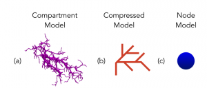
from https://doi.org/10.1101/177113
Surprisingly, they found that both the compressed geometry and single node models recapitulated key aspects of known network behavior, including the expected right-to-left motion stimulus response for four different T4 neuron types:
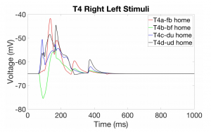
from https://doi.org/10.1101/177113
This data gets back to one of the questions posed by the Brain Emulation Roadmap, which is: what degree of complexity is required to model a particular function in a neural network stimulation?
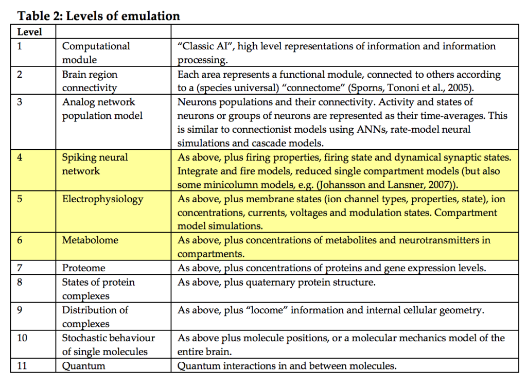
from http://www.fhi.ox.ac.uk/reports/2008%E2%80%903.pdf
Gornet et al.’s simulations fit somewhere between level #4 and #5 as defined by the roadmap authors.
Their results suggest that motion selectivity in T4 may only require a single compartment model for each neuron, rather than a model that allows for dendritic computation. But to emulate cognitive processes with complexity, more detailed neuronal and glial models will likely be required.
One limitation of their study that Gornet et al. note is that there are no detailed models of synapses, gap junctions, and neuromodulators in this brain region of Drosophila, so those parameters couldn’t be fully specified in their simulations, limiting their generalizability.
This is not just the case for Drosophila, as a lack of well-validated cellular models applies to nearly all model systems in neuroscience, and is a critical problem in need of a solution.
From a brain preservation perspective, this type of simulation work is critical because it helps us understand whether the information that we are preserving with a given brain preservation method will be sufficient to reproduce a particular function in a model system.

