
How much protein structure loss is there following glutaraldehyde crosslinking?
One of the key questions that I’ve had following the publication and success of aldehyde stabilized cryopreservation at mouse brain preservation is the degree to which the glutaraldehyde crosslinking step will lead to protein structure changes. In particular, I wondered about the prevalence of protein structure changes that would not allow inference about the original protein structure.
In December 2015, Murray et al. published a paper that helps address this. They invented a method for whole tissue preservation, SWITCH, which stands for System-Wide control of Interaction Time and kinetics of CHemicals.
One of the key steps of their process is glutaraldehyde crosslinking. In fact, they were able to control the timing of glutaraldehyde crosslinking by varying the pH of the system, since glutaraldehyde does not crosslink (due to protonated amine groups on proteins) at low pHs around < 3.
They first applied this technique to whole rat brains. They were able to get glutaraldehyde to diffuse efficiently throughout the brain by using a low-pH buffer, and then “turned on” its crosslinking with a neutral pH buffer, leading to a uniform glutaraldehyde-based hydrogel throughout the entire rat brain.
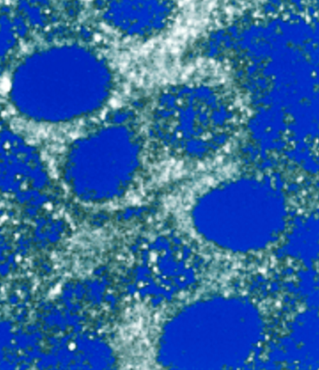
They then used a suite of antibodies to key neuronal proteins, including those that have been associated with memory, such as calbindin and NMDAR1.
They found that 86/90 of the antibodies that they tested were able to bind to proteins in the glutaraldehyde-fixed brain tissue, which was actually slightly better than the paraformaldehyde-fixed tissue, which had binding for 83/90 of the antibodies.
Small molecules such as dopamine, GABA, and neuropeptide Y were also able to be detected via antibodies (you can find the full list of molecules in Table S1 of their paper).
Thus, the authors were able to conclude that antigenicity is well maintained throughout the SWITCH preservation process. And since the SWITCH protocol prominently includes glutaraldehyde crosslinking, this gives me more confidence that glutaraldehyde crosslinking is unlikely to dramatically change the structure of key proteins and molecules in the brain.
Reference
Murray E, Cho JH, Goodwin D, et al. Simple, Scalable Proteomic Imaging for High-Dimensional Profiling of Intact Systems. Cell. 2015;163(6):1500-14.
Gazzaley AH, Weiland NG, Mcewen BS, Morrison JH. Differential regulation of NMDAR1 mRNA and protein by estradiol in the rat hippocampus. J Neurosci. 1996;16(21):6830-8.


